Chemo, Chemo, Chemo and She Died Within Two Years

Tin (M597) was 38 years old in 2007 when she found lumps in her breasts during a regular medical check up. A CT scan done on 21 May 2007 indicated: no lung nodules or abnormal masses or enlarged lymph nodes. In the left axilla are slightly prominent lymph nodes – these are under 1 cm in size. There are no liver lesions seen.

Tin underwent a left mastectomy on 1 June 2007.  The histology report indicated:

  1. Right breast biopsy – fibroadenoma in background of fibrocystic disease.
  2. Left breast (biopsy and mastectomy specimen): multifocal ductal carcinoma in-situ with invasive ductal carcinoma; invasive carcinoma measures 4.3 cm in maximum diameter; DCIS and invasive carcinoma extend close to the deep resection margin; skin and nipple are not involved by tumour.
  3. Left axillary lymph nodes: 4 out of 6 lymph nodes contain metastatic carcinoma, with extracapsular spread.
  4. TNM classification: pT2N2aMx, Stage 3A.

The tumour is negative for oestrogen and progesterone receptors, weakly reactive to C-erb-B2 and strongly reactive to P53. These results suggest that the breast cancer is unlikely to respond to tamoxifen / endocrine therapy.

As with most cancer patients, Tin underwent chemotherapy and radiotherapy after the surgery. Her husband was not sure how many courses of chemotherapy Tin had received. But one thing was sure – she was not getting any better. The oncologist behaved gently and encouraged Tin to continue taking the chemotherapy in spite of her deteriorating condition. She had full faith in her oncologist. According to her husband, Tin received Avastin towards the end of her chemo-treatment. Tin also underwent radiotherapy but her husband could not remember how many times she received the radiation treatment.

A follow up CT scan on 14 May 2008 indicated: no lung nodules or masses seen. Mediastinum remains clear of mediastinal lymphadenopathy. Liver also remains clear of metastatic deposits.

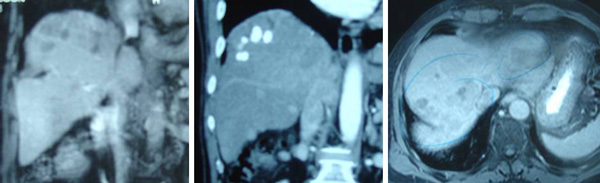
CT scan on 17 February 2009 indicated:

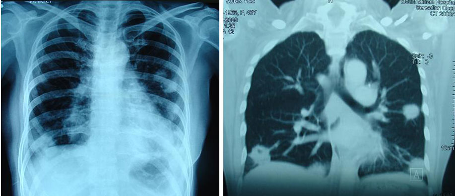
  1. Multiple pulmonary metastases are noted. This shows interval increase in size and number when compared with previous CT dated 22 December 2008.
  2. There is also interval development of bilateral pleural effusions, left larger than the right.
  3. Mediastinal and bilateral hilar lymphadenopathy are seen. This is unchanged when compared with previous CT. There is however interval development of left axillary nodes.
  4. There are now several small subcentimeter hypodense lesions seen in the liver compatible with hepatic metastases. These are not present in the previous CT.

CT scan on 8 June 2009 indicated:

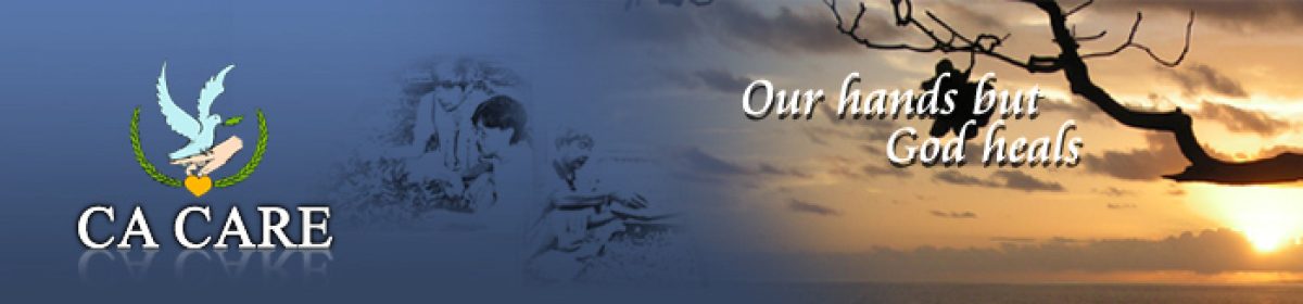
  1. There is extensive pleural shadowing seen in the lung fields on both sides with evidence of associated effusions noted.
  2. This is associated with lung nodules in both lungs the largest of which measures approximately 1 cm in the mid zone of the left side.
  3. A small associated pericardial effusion is seen.
  4. In the lower cuts, the liver outline shows no abnormalities.

Tin’s husband and mother-in-law came to seek our help on 2 July 2009. This is the gist of what we were told.

Tin’s husband and mother-in-law did not seem to agree that Tin undergo such extensive medical treatment but Tin insisted in carrying out what the oncologist wanted her to do. She questioned:Herbs are not proven, what if I took herbs and these do not work. As a result, her mother-in-law was afraid to give further comment. Even when Tin’s condition had turned worse and she became breathless due to fluid in her lungs and pericardium, the oncologist was saying there was nothing to worry. Every three weeks she had to have a blood transfusion. After Tin gets out of the hospital, the oncologist said she should come back to him for more chemotherapy.

Realizing that she was not going anywhere with medical treatment, Tin as last agreed to try our herbs. This resulted in Tin’s husband and mother-in-law flying to Penang to see us. We prescribed some herbs but expressed our skeptism.

On 19 July 2009, Tin’s husband came to Penang again. He informed us that Tin had been hospitalized and was only able to take the herbs for three days so far. Her condition was deteriorating. The doctor had requested the family to take Tin home. At most she would survive for another 2 months. Tin needed oxygen to help her breathe.

All in all, the family had spent approximately S$200,000 for the treatment. Tin’s husband told us: Please try to save my wife.

Barely 3 days after her husband’s second visit, we received this e-mail:

Wed, Jul 22, 2009 at 5:42 PM

Dear Mrs Teo,

Thank you so far for your help rendered by you and Dr. Teo. This morning at 9:21 am, my wife finally passed away after her struggle against the cancer for this past 2 years. She lost her battle ….

Comments: Is losing a battle against cancer such as this something unusual? Definitely not. At CA Care we witness see such disasters all too often. Tin had undergone all the medical treatments necessary for cancer and she died within two years. Where is the proven science in this case?

Granted, Tin’s cancer was serious – Stage 3A, but her CT scan of 14 May 2008 indicated no problem with her lungs or liver. There were also no serious problems with her lymph nodes. But one a half years later, trouble started brewing. CT scan in December 2008 indicated recurrence and it became even more serious in February 2009 in spite of more treatments (more of different poisons?).

By June 2009 Tin’s condition became more critical and she had fluid in her lungs and also her pericardium (heart lining). What had happened? Was this due to her cancer or the results of her treatment?

Often treatments of breast cancer are referred to as Slash, burn and poison. It is also said that these treatments are worse than the disease. What could have happened if Tin were to do nothing invasive and go for alternative treatment?  Having to suffer and to spend S$200,000 but ending up dead within 2 years is no bargain at all.

Dr. Frank Daudert of Pro Leben Klinik in Igls /Innsbruck, Austria said: Doctors give chemo, chemo, chemo. And patients die, die, die. He went on to say: Doctors are blindly giving chemotherapy to some patients while the cancer cells smile and the patients die.

Tin was totally committed to curing her cancer by the medical way. To her, herbalists are quacks and the herbs are not proven. She had resisted seeking alternative therapy or help. According to her family she only agreed to take the herbs in July 2009, and a few days later she died. That was a bit too late! There are many patients who are like her. Let this be a lesson to  numerous others who come after her.

Yee Died After Extensive and Costly Medical Treatments

Life on earth is a living experience. Let the death of Yee be a valuable lesson for many others who come after her.

Yee was 40 years old when she was diagnosed with breast cancer in October 2005. She underwent a mastectomy. It was a Stage 2 disease with no lymph node involvement. The tumour was 3 x 2 x 2 cm in size.

After surgery, Yee received 6 cycles of FAC chemotherapy (5-FU, Andiamycin and Cyclophosphamide). No radiotherapy was indicated. Chemotherapy was completed in March 2006 after that she was started on tamoxifen.

Yee was well for about 9 months. Sometime in January 2007, she noted a swelling in the right side of her neck. A CT scan of the thorax on 19 January 2007 indicated: multiple nodules scattered in both lung fields ranging from 2 to 10 mm. This means the cancer had spread to her lungs.

Yee was given 8 cycle of taxane-based chemotherapy. This treatment was not effective. CT scan on 4 July 2007 showed the nodules in the lungs were progressing.

Yee received more chemotherapy – 6 cycles of Navelbine + Herceptin. This treatment cost about RM 50,000. CT scan in November 2007 showed stable disease. From November 2007 till December 2008 Yee was on the oral drug, Tykerb (lapatinib) 4 to 5 tablets per day. Each tablet cost RM 65.00. At RM 260.00 per day this medication cost RM 7,800 per month. It seemed that the total cost for about 14 months on Tykerb came to about RM 93,000.00.

By February 2008, failures started to show up again. A CT scan on 13 February 2008 indicated:enlarging pulmonary nodules ranging from 0.5 to 2.2 cm.

Six months later, 19 August 2008, CT scan indicated pulmonary nodules are increasing in number and measure from 0.5 to 5.0 cm. Subcentimeter mediastinal nodes are also seen.

In October 2008, Yee suffered right arm pain and there was palpable swelling in the right collarbone. CT scan on 13 October 2008 showed more serious disease progression:

  1. fibrosis in the right axilla.
  2. an irregular ill-defined node in the right infraclavicular region, approximately 2.0 cm.
  3. another small right supraclavicular node, 0.7 cm is also present.
  4. medistinal nodes,size slightly increased from the last CT examination.
  5. pulmonary masses and nodules are enlarging. The largest mass, in the left lung is currently 5.6 cm. Other lesions measure from 1.0 to 4.8 cm.
  6. A new hypodense lesion, approximately 1.0 cm is now seen in Segment 7. This is suspicious of liver metastasis.

In view of the progressive disease, Yee underwent 28 times of radiation treatment starting 13 October 2008 until 1 December 2008, while at the same time continuing with her oral-drug, Tykerb.

On 17 December 2008, York suffered headaches and was unable to sleep. A CT scan done on 19 December 2008, indicated multiple brain metastases.

Yee and her husband came to seek our help on 5 December 2008. Yee’s condition was serious. Her right arm was numb. She had no appetite. She was severely breathless. Even the supply of oxygen to her nostrils did not help at all. She told her husband she would rather die. In early February 2009, Yee was admitted to the hospital where the doctor suggested that she receive radiation to her brain. It was not to be – she died even before the treatment.

This is a tragic case. Yee’s story is similar to that of Fransiska of Indonesia. Fransiska underwent surgery and received chemotherapy, radiotherapy, Herceptin and Tykerb + Xeloda. Fransiska died. She was diagnosed in November 2004 and died in December 2008. Yee was diagnosed with a Stage 2 breast cancer in October 2005, underwent similar treatments and she died in February 2009.

Doctors, the media and patients always look to newer drugs and newer technologies as a new hopefor treating illnesses. We have been hooked and made addicted to the idea that something new is always better. Herceptin and Tykerb are new bullets for cancer which are now beginning to appear in our local landscape. Are they better or dangerous?

From the website (http://www.tykerb.com ) you can learn that:

  1. There is no cure for metastatic breast cancer. But it can be treated. What do you by such a vague treatment? Surely we can treat anything if there is money!
  2. Some women may develop liver damage while taking Tykerb. The cause of this damage is not known. Liver damage may be severe and may cause death.
  3. The most common side effects of Tykerb are diarrhea; vomiting; feeling sick to your stomach; feeling tired; red, painful hands and feet; and rash.
  4. Dry cough or have shortness of breath may be signs of inflammation in the lungs.

Can Herceptin cause any serious side effects? Yes, according to the National Cancer Institute website (http://www.cancer.gov/cancertopics/factsheet/therapy/herceptin)

1.       Herceptin can cause heart muscle damage that can lead to heart failure.

2.       Herceptin can also affect the lungs, causing severe or life-threatening breathing problems.

3.       Herceptin can cause allergic reactions that can be severe or life-threatening. Symptoms of a reaction include a drop in blood pressure, shortness of breath, rashes and wheezing.

4.       Because of these potentially life-threatening side effects, doctors are WARNED to evaluate patients carefully for any heart or lung problems before starting treatment. Do you think they ever do this? In this case, Yee had metastasis (that is not severe problem enough?) to her lungs. As such would Herceptin not make things worse for her? When she came to us she was severely breathless. What could have been the cause?

And one most important question which patients (and doctors?) do not even care to ask: Does Herceptin cure breast cancer? The answer is absolutely NO. Addition of Herceptin to the normal chemotherapy regime prolonged survival by 4.6 months.  Nowhere in the website ever says that Herceptin cures breast cancer. Read the section: Possible Benefits of Herceptin (take note, even the title itself does not seem to inspire much confidence and hope) in its official website: http://www.herceptin.com/adjuvant/what-is/benefits.jsp

Dr. Ralph Moss wrote a report entitled: Herceptin or Deception. Michael Janson, M.D., past president of the American College of Advancement of Medicine gave this comment about the report: Dr. Mossreveals the hollow core of the recent medical reports on Herceptin, showing that it is not what has been claimed, and that the statistics were manipulated to make it seem far better that it is, while underplaying the potential risks.

In Yee’s case, like in many other cases before her, perhaps there is no point asking the same question: What has gone wrong? Things seem to go wrong most of the time. Allow me to think aloud:How could a Stage 2 breast cancer kill a patient within four years? Would she have died if she was to do nothing?

Breast cancer is less common among those in their thirties, but if breast cancer occurs in younger women, it tends to be more aggressive than in older women. While this may be true, some people want to make us believe that it is because Yee was young and had an aggressive type of cancer that make her treatment ineffective. Let me invite you read the case of three ladies who were before forty when they had cancer yet they survive. The only thing they did differently was that they did not blindly their doctors – they declined chemotherapy, radiotherapy or hormonal treatments.

Tee, a 38-year-old lady who was diagnosed with breast cancer in October 2005 (note: at about the same time as Yee) and she refused chemotherapy. Tee is still alive as of this writing (March 2009) while Yee died in February 2009. You can read Tee’s story in our Case Report series.

Sue was 39 years old when she discovered 2 lumps in her right breast in 2003. She underwent a mastectomy. Her oncologist told her that with chemotherapy she would have 5% chance more of survival. She but declined further medical treatment, including tamoxifen as suggested by her surgeon. Sue change her diet and lifestyle and took herbs. It has been 6 years now and Sue has been leading a trouble-free life.

Julia discovered a 4 cm lump in her breast in 1995. She was then 36 years old. She was asked to undergo a mastectomy. She declined and never went to see her doctor again. She declined chemotherapy, radiotherapy or hormonal therapy and decided to seek alternative healing. She father is an herbalist and naturally she turned to him for help. Julia’s healing journey along the uncharted path is interesting and sometime dangerous. But to fact remains she is still very much alive and well as of this writing (2009). It was 14 years that she was first diagnosed with cancer. The detail story of Julia is found in our book: The herbal option, Chapter 3.

Cindy was 34-years old when she was diagnosed with breast cancer in August 1994. She underwent a lumpectomy. Since the margin was not clear she was asked to undergo a mastectomy to be followed by chemotherapy and radiotherapy. She declined further medical treatment. She changed her lifestyle and diet, did qi gong and meditation and sought homeopathic therapy. It has been 15 years and Cindy has been leading a healthy without any problem (Chapter 2: The herbal option).

Dr. Gershom Zajicek, Professor of Medicine at The Hubert H. Humphrey Center For Experimental Medicine and Cancer Research, The Hebrew University of Jerusalem, Israel (http://www.what-is-cancer.com) wrote: Modern medicine has the best means to treat disease, yet the basic tenets of treatment are false.

Dr. Frank Daudert, Pro Leben Klinik in Austria said: Doctors are blindly giving chemotherapy  … while the cancer cells smile. Doctors give chemo, chemo, chemo. And patients die, die, die.

In their book: More harm than good, Alan Zelicoff, M.D., and Michael Bellomo, J.D./MBA, wrote: (Physicians are) taught that doing nothing almost guarantees that the patient will suffer and that doing something outweighs doing nothing. The truth is that the vast majority of cancer – once spread – remain incurable despite the availability of many dozens of new chemotherapeutic drugs and even the use of antibodies directed against cancer cells.

Richard Deyo and Donald Patrick, professors, University of Washington, Seattle, USA, wrote in their book: Hope or hype – the obsession with medical advances and the high cost of false promises: We are born with our own blind trust in a medical establishment that preys on our deepest fears, all the while purporting to ride to our rescue with “miracles cures.” Indeed many medical advances do offer real advantages but there are as many others that offer little, if any, advantage and many have alarming side effects … marginally effective at best – and sometimes downright dangerous …. They often lead to useless, harmful and unnecessarily expensive care … When doctors introduce new products good money often trumps good science.


Surgery, Chemotherapy, Radiotherapy and Hormonal and Targeted Therapy Did Not Cure Breast Cancer

Case 1

Fay (not real name) is a 45-year-old Malaysian. She was diagnosed with breast cancer in September 2006.

  • She underwent a mastectomy with axillary clearance (removal of lymph nodes).
  • After the surgery she received 25 radiation treatments and six cycles of chemotherapy. The drugs used were 5-FU, epirubin and cyclophosphamide (FEC). All treatments were completed in April 2007.
  • Every 4 months Fay went back to her oncologist for routine surveillance check up. Everything was alright.
  • In August 2008, cancer was found in her bones – L2, L5, sacrum and pelvis.
  • She had been on tamoxifen for almost 2 years (November 2006 to August 2008). Tamoxifen had failed and the doctor suggested that she take another drugs, Arimidex.
  • She received to receive more chemotherapy and suffered badly from the side effects.

Case 2

Rin (not real name) and is a 40-year-old Indonesian lady living in the United States. She wrote:

  • First I was diagnosed with breast cancer in December 2004.
  • I did the lumpectomy on the left breast in February 2005.
  • After the surgery, I received 8 cycles of chemotherapy. After the chemotherapy I had menopause.
  • I then had radiotherapy for 35 times and I finished it in October 2005.
  • I took tamoxifen, 20 mg once a day.
  • I did regular check up with my oncologist every 6 month and I did yearly mammogram and the past 2 years I also did the bone density test.
  • In August 2008, I started feeling pain in my left leg and sometimes in my left arm. The pain did not go away and it hurt more and more. Then I was not able to and walk straight and could not bend. It was very hard for me to go up and down the stairs.
  • In November 2008, I had a whole body scan and also CT scan. The cancer had spread to the bones –  left humeral head, left femur and L5.
  • I again did radiotherapy for the effected area for 10 times.
  • In Dec 2008, I developed blood clots in my left leg.
  • My doctor changed my medication from tamoxifen to Arimidex.

Case 3

Gay (not real name) is a 43-year-old Australian. She was diagnosed with breast cancer in 1999. She wrote:

  • I received 6 months of chemothrapy and 3 months radiation therapy.
  • Then I was started on tamoxifen for 5 years followed by Arimidex.
  • I have had no problems until the past 6 months. I had a slight pain in my right upper abdominal. My tumor markers were elevated.
  • After a number of scans, it turned out to be bone metastases.

Case 4

Sri (not real name), 57-year-old Indonesian, was diagnosed with left breast cancer in 2003. She underwent a mastectomy followed by chemotherapy and radiotherapy. At the time we talked to her, Sri appeared to have chemo brain and was unable to recollect the details of her treatment. Her response to our questions was also very slow. Sri received all these treatments in New Zealand.

Sri went back to her doctor for routine check up and was told everything was fine. However, in 2007, she did not feel well. Further examination indicated bone metastases. She again received six cycles of chemotherapy and 10 radiation treatments. All treatments were completed in November 2008. Sri came to Penang in February 2009 and did a CT scan. The results indicated the following:

  • Lesion in T1 and T5 vertebrae.
  • Nodule in C5 and lesion in L4 vertebral bodies.
  • Several lytic lesions in the left iliac bones.
  • Suggestive of liver cirrhosis.

What can we learn from these four cases?

1. These patients had undergone all the required medical treatments – surgery, chemotherapy, radiotherapy, and oral drugs – tamoxifen and Arimidex. They had received the best that medical science has to offer but cancer still recurs.

2. Oncologists say all these are treatments are scientifically proven, FDA-approved backed by data reported in peer-reviewed medical journals. The questions are: What is so great about all these? Why do these patients still suffer metastasis? What is the “real and honest” truth about all these treatments?

3. Does it ever cross somebody’s mind that the inability to cure or the ability of the cancer to spread could also be due to the treatments themselves?

4. Look at these cases again. Fay in Malaysia suffered metastasis 1 year and 4 months after completion of her medical treatment. Rin from USA and Sri in New Zealand suffered metastasis barely three years after her treatment while Gay from Australia had metastasis about eight years after her treatment. These cases reflect similar problems faced by most patients everywhere in the world. It does not matter where you live and who or what you are, doing the same thing produces the same results.

Einstein said: Insanity is to the do the same thing over and over again and expecting different results. Can you learn anything from the wisdom of this great Man of Science? Patients in the USA,Australia, New Zealand and Malaysia received the same kind of treatments and all of them ended up with the same results. Over the years, I have observed similar stories being repeated over and over again so much so that bony metastasis can or is expected after such treatments.  To expect otherwise is what Einstein said is insanity.

The questions that boggle the mind are: Why are those in the know NOT doing anything about it?Why are patients kept in the dark and not warned about this possibility? Perhaps we can do MORE than just prescribing drugs? Tamoxifen is supposed to prevent recurrence but in all the cases above, it has failed miserably. Why don’t we look beyond what are being done today?


Liver Cancer: Surgery and Chemo Did Not Cure Him

Surgeon told him three years too late that nine out of ten patients whom he operated on died anyway

KB (H467) is a 57-year-old male. In 2006, he suffered back pains. CT scan of the abdomen on 23 June 2006 indicated: “a hypervascular lesion in the right lobe of liver in Segment 6/7 measuring 3.5 x 5.2 x 4.5 cm. This has ill-defined margins. Features suggestive of an underlying hepatoma.”  In short, KB had liver cancer and without hesitation and in haste due to fear, he underwent liver surgery in a private hospital in Penang. This cost him RM35,000.

As a follow-up treatment, KB underwent seven times of  chemoembolisation. Each treatment cost RM5,000. The first five treatments were well tolerated but the last two caused severe side effects. It was a difficult experience and KB said he was “half dead”.

About three years later, KB felt a lump in his abdomen. CT scan on 23 June 2009 report is a follows:

“There is a previous segment 6/7 and 7/8 wedge liver resections. There are multiple foci of lipiodol uptake in the liver post chemoembolisation in keeping with multicentric hepatomas.  These range from 2 – 20 mm in size. There are several recurrent hypervascular foci noted scattered in the subdiaphragmatic region of the right lobe of liver ranging from 5-15 mm. Absent gallbladder (previous cholecystectomy). No evidence of abdominal lymphadenopathy.”

Another CT scan was done on 4 September 2009. The imaging report appeared exactly the same as the one written on 23 June 2009 with the following exception:

“There are several hypodense foci in the right subdiaphragmatic region ranging from 2-15 mm. Impression: several  hypodense lesions scattered in the subduaphragmatic region of the right lobe of liver, ? generating nodules, ? early hepatomas.

MRI was also done on 3 October 2009. The report appeared exactly the same as the CT scan reports of 23 June and 4 September 2010, with the following exception:

“There are multiple irregularly enhancing nodules scattered in both lobes of the liver with clustering in the right subdiaphragmatic regions ranging from 3- 20 mm. These are suspicious of multicentric hepatomas.”

It was a real disappointing result for KB and his family. At that time he was still undergoing chemoembolisation. KB complained to the surgeon that the treatment was making him sick. The surgeon said this: “Out of ten patients I operated on, nine died.” KB and his family were also told that KB was already lucky that he is still alive. “Most of others patients have all died. If you do not wish to continue with the chemo, you would be waiting to die.”

Listen to our video conversation below.

The son told us: But he only told us this fact three years after the operation. The surgeon should have told us before he did the operation.  Disappointed KB decided to see another doctor in Ipoh. A total body CT scan was done on 10 August 2010. The report is a follows:

“There are multiple hypodense lesions seen within the liver affecting nearly the entire parenchyma, with clustering seen in the subdiaphagramatic region. This is suggestive of multicentric hepatoma. The area of involvement is worse than that seen in previous scans.

In the private hospital in Ipoh, KB received three i/v chemo treatments. Unfortunately the treatment was not effective.  The family was told about CA Care and KB and his family came to see us on 26 November 2010.  Take a look as his blood test results.

  4 Oct 10 15 Oct 10 4 Nov 10 19 Nov 10
Direct bilirubin 7.3 6.7 5.9 7.4
SGOT/AST 116 103 112 132
SGPT/ALT 66 49 55 74
GGT 882 723 649 790
Alkaline phosphatase 179 157 138 179
Alpha-fetoprotein 2480 2419 3185 3208

Comments:

This is indeed a sad story. Talking to KB and members of his family that night, made me realized how vulnerable we all are. KB trusted the expert to cure him. He was willing to spend so much money to find the cure for his liver cancer. He did not doubt about what he was doing  – for that matter he probably took it for granted that he would get his cure. Like most people believe, after the operation the cancer is gone.

Then came the bombshell three years too late. He was told that nine out of ten patients who underwent liver surgery died anyway! I asked him: “Before you had the surgery, did you ever ask the doctor if surgery would cure you?” The answer was NO. Well, most patients never ask such a question. They took for granted that surgery would be the “magic tool”. His wife said: “We were ignorant”. KB said: “I was in fear.”

Dr. Hamilton, a neurosurgeon, in his book: The scalpel and the soul, wrote: “As doctors, we generally don’t tell outright lies. We just don’t speak the truth fully.”

So, here it is. If you don’t ask you get no answer. There is a Malay saying: Segan bertanya, sesat jalan (Shy to ask, you lose your way).

This is what I always tell patients. Before you do anything – be it surgery, chemotherapy, radiotherapy or even taking drugs – ask the doctor first of the possible and expected outcome. Why the rush to get things done? Ask if what he is going to do or giving you is going to cure you or not. Then evaluate his answer or response. What he says, how he says it and how he addresses your concern (including his body language) will tell you where you are heading for.

I was a bit baffled that night. I asked KB and his family – what made the surgeon say that damaging “confession” – nine out of ten patients whom he operated on died. In what context or under what circumstance did he say such a thing? I got this answer. KB was unhappy because the chemo was “killing” him and he complained to the surgeon about it. He was reluctant to go on further with the chemo – in spite of having received seven “shots” already. In an effort to make KB comply with his instruction, perhaps “threatening” KB with a possible death would do the trick?

But then why come out with such damaging statement? One is tempted to speculate that this “truth” has been lying buried in his heart all these days of his professional life but he could not share this truth with his patients. But once a while, things have to “explode” and be let out. Unfortunately for KB, this truth is revealed three years too late. What if KB was told that he had only a 10 percent chance of survival before he underwent the surgery? Would he go ahead with the surgery?

Why must doctors adopt the attitude that “we just don’t speak the truth fully”?

At CA Care we lay out our cards on the table – clearly and fully. When KB came to see us, I have made it clear that I would not be able to cure him at all. But I promise to do my best to help him – the way that I know how. I told him to keep to the good diet. Cancer patients cannot eat anything they like. I am aware that most patients are disappointed and they never come back again after hearing my “lecture.” That is okay – it is better to be honest and become unpopular with patients than misleading them.  The wise words of Rabbi Harold Kushner came to mind. In this world we have a choice on how we conduct our affairs:  “There is the morality of cleverness and wit, in which success means getting the better of the other person by means of a slick business deal or a clever answer. Then there is the morality of righteousness, in which the highest good is thoughtfulness toward others. The worst sin is hurting another person.”

In his book, Never to be lied to again, Dr. David Lieberman wrote:

  • In an ideal society there would be no need for lies. But we live in a world of deception. And whether you want to play or not, you’re in the game.
  • And there’s one undeniable truth about lying – everybody does it.
  • Some of these lies can be “subtle that they can be easily missed unless you pay close attention”. Others may be glaringly obvious. In some instances, you’ll be looking for lies of omission – what’s missing that should be there.

Dr. Lieberman offered numerous tips in his book on how to detect lies and deception. Here are some examples:

  • Body language – the truth can be always silently observed. No or little direct eye contact is a classic sign of deception. Feeling guilty a lier does not want to look you in the eyes. Liers tend to be less expressive with his hands or arms. Arm movements and gestures seem stiff and almost mechanical – not natural.
  • Someone who is lying rarely points a finger, either at others or straight up in the air. Finger pointing indicates conviction and authority.
  • Watch out for the good old Freudian slip or slip of the tongue.  This is a subconscious leak when a person’s misspoken words reflect and reveal his true feelings, thought or intentions.
  • He doesn’t want to respond to a question. He may imply an answer but never answers  it directly.
  • When asked a question, he depersonalizes his answer by offering his belief on the subject instead of answering directly.

Here are something we can do and need to be aware of:

  • When you’re desperate, you’ll be apt to do what you never should do – make a decision out of fear.
  • Just because you’re told that something is the latest, best, hottest or biggest seller does not make it right for you.
  • A white lab coat does not make anyone an expert.
  • Evaluate a person’s integrity based upon what is being presented not what is promised.
  • Often we don’t stop and ask ourselves, “Does this make sense?” A dash of common sense can go a long, long way.
  • We certainly can’t stop people from trying to lie to us, but we can keep them from being successful.

Read the following related posts:

A Great Failure and Let Down

Surgery for Liver Cancer: Eighty Percent Success?

Liver Cancer: He Died After a RM 10,000 Open-Close Surgery

The Doctor Said: “No More Hope” After a 60-Million-Rupiah Operation for Bile Duct Cancer

Healing of Metastatic Liver Cancer


The Doctor Said: “No More Hope” After a 60-Million-Rupiah Operation for Bile Duct Cancer

Hass (not real name, M847) is a 44-year-old male from Indonesia.  In early 2010, he had jaundice. The doctor suspected this was due to Hepatitis. Hass was prescribed medication. After a month, the treatment was not effective. Hass went to a private hospital in Kuching.

A CT scan on 28 January 2010 indicated a 6 mm stone in the gallbladder. There was a 1 cm hypodense lesion in segment 8 of his liver. This could be just a cyst.  The common bile duct was dilated. The maximum diameter was 16 mm. There was a 18 mm mass at the distal common duct. Conclusion: Mass in the lower common duct with biliary obstruction.

Immediately Hass underwent an operation to remove his gallbladder and the bile duct tumour. The histopathology report dated 3 February 2010 indicated: infiltrating moderately differentiated exophytic adenocarcinoma of bile duct, invading through entire thickness of the wall and metastatic to periductal lymph nodes. Margins of excision free of tumour.

An ultrasound of the abdomen on 8 June 2010 showed no evidence of tumour recurrence although his CA19.9 increased from 52.4 (in March 2010) to 1420.0. A CT scan on 9 June 2010 indicated a 15 x 10mm lesion in segment 7 of his liver. The 10 mm cyst in segment 8 of his liver remained the same. In addition there were a few paraaortic lymph nodes noted from the celiac axis down to the liver of the renal hilum. The largest node measured 20.0 mm – the others were subcentimeter.

The doctor told Hass that the cancer had recurred and he was unable to help him anymore. Hass was asked to undergo chemotherapy. But he refused.

Below is a table showing the rise of CA 19.9 markers from March to October 2010.

Date CA 19.9
Before operation 2,000 plus
18 March 2010 – after operation 52.5
8 June 2010 1,420.0
3 August 2010 – last visit to surgeon 5,645.0
1 October 2010 18,746.18

Hass came to see us in Penang on 8 October 2010. Listen to our conversation.

Review of Medical Literature

Understanding the Liver-Gallbladder-Pancreas-Bile Duct Complex

  • Attached to the liver, is a pear-shaped structure called the gallbladder.
  • The liver makes bile and this is transported to the intestine by a long tube-like structure called the bile duct.
  • The gallbladder is attached to the bile duct by a small tube called the cystic duct.
  • The top half of the bile duct is associated with the liver while the bottom half of the bile duct is associated the pancreas through which it passes on its way to the intestine.
  • Blockage of the bile duct by a cancer prevents the bile from being transported to the intestine. When this happens the bile accumulates in the blood giving rise to jaundice. The skin and eyes becomes yellow. This condition may also cause severe itchiness.

(Diagrams are obtained from the internet)

The clinical presentation, prognosis and natural history of bile duct cancer (or cholangiocarcinoma) depend on where the cancer originates. The cancer may occur:

  • In the part of the bile ducts contained within the liver. This is known as intra-hepatic.
  • In the part of the bile ducts outside the liver. This is known as extra-hepatic.
  • In the duct between the left and right portal veins. This is known as hilar bifurcation.

Anderson et al. in Diagnosis and treatment of cholangiocarcinoma, The Oncologist, Vol. 9, No. 1, 43–57, February 2004, noted that approximately:

  • 60%-70% of cholangiocarcinomas occur at the hilar bifurcation.
  • 20%-30% in the distal common bile duct.
  • 5%-15% within the liver (intra-hepatic).

Patients with extrahepatic tumors usually present with painless jaundice due to biliary obstruction, while patients with intrahepatic tumors usually present with pains, pruritus (66%), abdominal pain (30%-50%), weight loss (30%-50%), and fever (up to 20%).

In their review, Anderson et al also noted that:

  • The majority of patients with cholangiocarcinoma presents with unresectable,advanced stage disease and have a survival of less than 12 months. Cure rates are low, even with aggressive therapy. The average 5-year survival rate is 5%-10%.
  • Surgery remains the only intervention offering the possibility of a cure. But the treatment goal should be complete excision with negative margins.
  • All patients should be fully evaluated for resectability before any type of intervention is performed.
  • Neoadjuvant and adjuvant therapies have not improved survival in patients with this tumor.
  • Since the prognosis for unresectable patients is poor, palliative measures should be aimed at increasing quality of life first and increasing survival second.

Results of Surgery

  • Among selected patients who undergo potentially curative resections, 5-year survival rates are generally from 8%-44%.
  • The importance of achieving a margin-negative resection cannot be overemphasized. The 5-year survival rates were greater when a negative margin was obtained, 19%-47% versus 0%-12%.
  • Distal lesions represent approximately 20%-30% of all cholangiocarcinomas and are usually treated with pancreaticoduodenectomy (Whipple procedure). Achieving a margin-negative resection is also important with these tumors. The 5-year survival rates in selected patients who underwent curative resections is 21%-54%.
  • Intrahepatic cholangiocarcinoma is usually treated by hepatic resection. Three-year survival rate of 60% was reported in patients who underwent a margin-negative partial hepatectomy.

Chemotherapy

  • Chemotherapy has not been shown to markedly improve survival in patients with either resected or unresected cholangiocarcinoma. The majority of reports use 5-FU alone or in combination with methotrexate, leucovorin, cisplatin, mitomycin C, or interferon alpha.
  • The 5-year survival rates were not significantly different between patients who received chemotherapy and surgery and those who received surgery alone.

A note in http://emedicine.medscape.com/article/189843-treatment stated  that:  “Chemotherapy has not been proven to be of definite benefit.”

Experience of doctors in Fuda Hospital, China indicated that:

  • Cancer arising in the extrahepatic bile duct is an uncommon disease, curable by surgery in fewer than 10% of all cases.
  • In most patients, the tumor cannot be completely removed by surgery and is incurable.
  • Palliative resections or other palliative measures e.g. stenting procedures may maintain adequate biliary drainage and allow for improved survival.

Experience of doctors in India indicated that:

  • Surgery may be used to remove the cancer if it has not spread beyond the bile duct. It is not always possible to carry out surgery, as the bile duct is in a difficult position and it may be impossible to remove the cancer completely.
  • There are different surgical procedures depending upon how big the cancer is and whether it has begun to spread into nearby tissues.

These surgical procedures are:

  • Removal of the bile ducts. If the cancer is small and contained within the ducts, then just the bile ducts containing the cancer are removed.
  • Partial liver resection. If the cancer has begun to spread into the liver, the affected part of the liver is removed, along with the bile ducts.
  • Whipple’s. If the cancer is larger and has spread into nearby structures, then the bile ducts, part of the stomach, part of the duodenum, the pancreas, gall bladder and the surrounding lymph nodes are all removed.
  • Bypass surgery. Sometimes it is not possible to remove the tumour. Other procedures may be performed to relieve the blockage and allow the bile to go into the intestine. The jaundice will then clear up.
  • Stent insertion. One way to relieve jaundice without a surgical operation is to perform ERCP (Endoscopic Retrograde Cholangiopancreatography). This procedure involves insertion of a tube, called a stent into the blocked bile duct. The stent clears a passage through the bile duct to allow the bile to drain away. The tube usually needs to be replaced every three to four months to prevent it becoming blocked. If the tube does block, recurrent high temperatures and/or return of the jaundice will occur.

In summary, Witzigmann et al. in an article, Guidelines for palliative surgery of cholangiocarcinoma, HPB (Oxford). 2008 June 1; 10(3): 154–160, listed the following guidelines for surgery of bile duct cancer:

  1. Non-surgical stenting is regarded as the first choice of palliation for patients with short life expectancy.
  2. For patients with longer projected survival, surgical bypass should be considered.
  3. Palliative resections may be beneficial to patients with distal and hilar bifurcation.

Questions to Ponder

The fact is very clear. Surgery for bile duct cancer in this case is an absolute failure. Is this failure a rare event? Probably NOT! A surgeon told his liver patient: “For ten liver operations that I performed, nine patients died”. Good information indeed but the problem is that he made this comment some three years too late. This patient had already undergone the operation and had suffered a severe recurrence.

A few more questions come to mind:

  • According to the literature, not all patients with bile duct cancer are suitable condidates for surgery.  “All patients should be fully evaluated for resectability before any type of intervention is performed”. To what extent is this evaluation done?
  • The surgeon recommended chemotherapy after the recurrence. What benefit can the patient expect from this? Medical literature clearly indicates that “Chemotherapy has not been proven to be of definite benefit.”
  • Is the suggestion of chemotherapy after the failure of surgery, a way of “passing the buck” to someone else? “I have done my job to cut it out. My responsibility is over. Now you go to the oncologist and see what he can do for you.”

More related stories:

Liver Cancer: Surgery and Chemo Did Not Cure Him

A Great Failure and Let Down

Surgery for Liver Cancer: Eighty Percent Success?

Liver Cancer: He Died After a RM 10,000 Open-Close Surgery

Healing of Metastatic Liver Cancer