Mediastinal Germ Cell Tumour: Nonosrugery, Chemotherapy and Hyperthermia Failed to Cure Him

Ali (not real name) is 24 years old. This is his story.

In November 2011, Ali noticed “floating” veins in his chest. His face was swollen. He was admitted to a private hospital. A CT scan showed a mass in his chest. A biopsy confirmed a mediastinal germ cell tumour.

Ali subsequently undergo 4 cycles of chemotherapy using BEP regimen (Bleomycin + Etoposide + Cisplatin).

His AFP (alpha-fetoprotein) decreased following the treatment. In spite of that, the surgeon was unable to operate on Ali because the tumour was found to grow around his heart. Instead of surgery, Ali had to do 30 sessions of radiotherapy. Unfortunately, radiotherapy did not help much.

In March 2014, Ali went to London to undergo a nanosurgery. According to the English doctor, this procedure would help Ali. Indeed after the surgery, the AFP decreased, but later the AFP increased again.

Ali returned to Malaysia and underwent another 4 cycles of chemotherapy using TIP (paclitaxel + ifofamide + cisplatin). TIP regimen is a second-line salvage chemotherapy for patients post-BEP.  His AFP decreased after TIP but the size of the tumour remained the same — 4.3 cm. The tumour size before any treatment was about 8 cm.

Ali consulted another oncologist. Since nothing much could be done, the doctor recommended that Ali go to Germany for hyperthermia + chemotherapy treatments. Ali stayed in Germany for about a month undergoing that treatment which cost about RM 80,000.

Ali returned to Malaysia and continued with the hyperthermia and high dose chemo. He had been on this treatment for almost a year now. His AFP decreased but the size of the tumour stubbornly remained the same.

Ali again started with another round of chemotherapy using Avastin + Gemzar + Taxol and Oxaliplatin. This treatment made Ali real sick. He needed a blood transfusion. In addition, he started to cough out blood. His AFP shot up to 24,591, as on 5 February 2015.

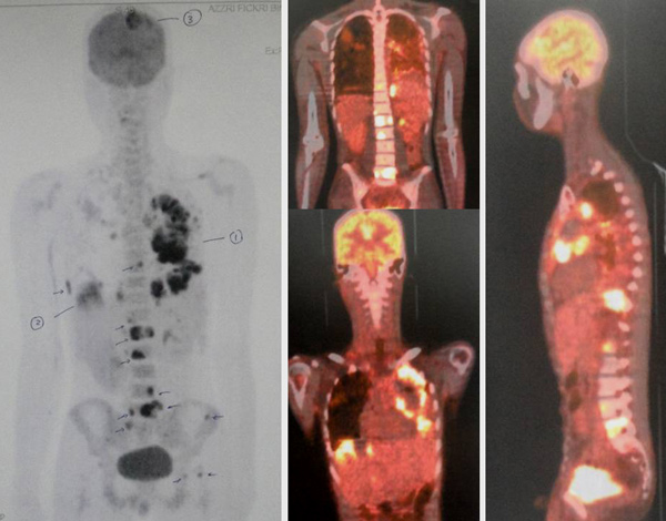
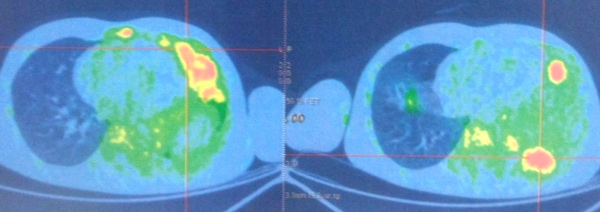
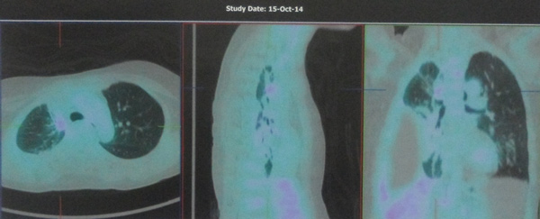
A PET scan on 6 February 2015 indicated:

  • extensive malignant lesions present in the left lung, less in the right lung.
  • several metastases present in the bones.
  • a large nodule about 4.5 cm in the right lobe of his liver and a few small ones.
  • a lesion about 4.0 cm in the parietal region of his brain.

Composite-1-overall2-Brain 3-Lungs

In March 2015, Ali and his parents came to seek our opinion.

I felt empathy sitting in front of this young man listening to his sad story. I admitted that there is nothing much I could do. It would be wrong to believe that our herbs could solve his problem. Even more so, after all that modern medicine could offer had failed.

In writing this story, I am reminded of what Drs. Jerome Groopman and Pamela Hartzband wrote in their book, Your Medical Mind about the many types of patient’s mindset.

Naturalism vs Technology

  • Some are naturalism orientated believing that the body can often heal itself if given the proper environment, harnessing the mind-body connection and supplementing with herbs, vitamins, and other natural products.
  • On the opposite end of the this spectrum is the technology orientated believing that cutting-edge research yielding new medications and innovative procedures holds the answers. 

Maximal vs Minimal Treatment

  • Some people are profoundly proactive about their health, believing that more is usually better.
  • In contrast, those with a minimalist mind-set aim to avoid treatment if at all possible, and if not possible, they try to use the fewest medications at the lowest possible doses or to select the most conservative surgery or procedure. Minimalists hold to the notion that “less is more. 

Believers vs Doubters

  • Believers approach their options with the sense that there is a successful solution for their problem somewhere.
  • Doubters approach all treatment options with profound skepticism. They are deeply risk-averse, acutely aware of the potential side effects and limitations of drugs and procedures.

If patients come to see us hoping to find some kind of magic bullet, then it would be most disappointing to know that we have none. We try to help you the best we know how but we need your commitment to help yourself first. You need to have the right kind of mindset first.

It was indeed sad to note that he had submitted to the last round of chemo with Avastin + Gemzar + Taxol and Oxaliplatin., resulting him to cough out blood. One wonder, what did you expect to get from such treatment when earlier treatment did not work? Then, the bleeding. What caused this?

Click this link: http://www.avastin.com/patient. Most serious side effects: GI perforation: A hole that develops in your stomach or intestine. Symptoms include pain in your abdomen, nausea, vomiting, constipation, or fever. Wounds that don’t heal: A cut made during surgery can be slow to heal or may not fully heal. Avastin should not be used for at least 28 days before or after surgery and until surgical wounds are fully healed. Serious bleeding: This includes vomiting or coughing up blood; bleeding in the stomach, brain, or spinal cord; nosebleeds; and vaginal bleeding. If you recently coughed up blood or had serious bleeding, be sure to tell your doctor.

BY DOCUMENTING THIS STORY IT IS OUR HOPE THAT YOU OR YOUR LOVED ONES CAN LEARN SOMETHING MORE THAN JUST “GOING TO THE DOCTOR OR HEALER” TO SEEK TREATMENT.

2.3 cm Malignant Breast Lump: Surgery, Chemo and Radiation — Disaster

This is a tragic story which I find it real hard to “understand.” WF is 32 years old. In early 2014, WF felt a lump in her left breast. At that time she was pregnant and was about to deliver her baby. So nothing was done until after the birth of her baby.

On 14 March 2014, WF had an ultrasound of her breasts. “There is a 17 mm x 9.6 mm lesion at 2 o’clock position of left breast, 4 cm from the nipple.” A FNAC (Fine needle aspiration cytology) done in a Taiping private hospital showed “benign breast lesion.”

WF did another FNAC in April 2014. This time it was done in a private hospital in Penang. Unfortunately, the result showed “atypical cells … Highly suspicious of an infiltrating duct carcinoma.”

A trucut biopsy was done on 12 April 2014 confirmed an invasive ductal carcinoma.

WF consulted another doctor in another private hospital.

25 April 2014Ultrasound of Both Breasts Irregular hypoechoic lesion between 1-2 o’clock. It measures 23 x 18 x 12 mm. Some microcalcifications seen. In keeping with a neoplasic lesion.

Based on the above, WF had surgery. A wide local excision of the left breast mass was done (lumpectomy). The tumour removed was 23 mm in size. Two of the axillary lymph nodes were involved. All resection margins were free of malignancy. Immunohistochemical study indicated a triple negative tumour: ER negative, PR negative and c-erb-B2 negative. It was a Stage 2B cancer.

9 May 2014Ultrasound of Thyroid Multiple tiny nodules seen on both thyroid lobes, likely benign.

WF subsequently had 6 cycles of chemotherapy. Neither she nor her husband knew what drugs were used. Anyway, each cycle cost RM 6,000. WF lost her hair, felt tired and nauseous during her treatment. Chemotherapy was completed by October 2014. Then WF received 20 sessions of radiation and this was completed in November 2014.

About a month later, in late December 2014, the cancer spread to WF’s brain. There were 3 lesions in her brain. WF received 2 sessions of radiation to her head in January 2015.

Two months later, March 2015, CT scan showed the cancer had spread to her lungs, bone and liver.

WF was again asked to undergo 4 cycles of chemotherapy. WF did one cycle after which she and her husband came to see us and decided not to proceed with the treatment.

Chris: Did you ever ask the doctor if surgery, chemo and radiation were going to cure your cancer?

Husband: The doctors said there is a  80 percent chance of cure?

Chris: Did you ever ask what happen to the remaining 20 percent?

No reply.

Study the numbers of her blood tests.

Date CEA CA 15.3 (normal 0-32)
5 June 2014 Less 0.5 12.3
18 Nov 2014 0.4 9.7
10 Feb 2015 Less 0.5 13.2
10 March 2015 n/a 20.3
24 March 2015 n/a 37.0
7 April 2015 n/a 96.1
22 April 2015 1.4 142.6

In March 2015, WF was started on chemotherapy again because her CA 15.3 started to rise, indicating that the earlier chemotherapy had failed. Therefore, the answer is more and more chemo?

The following are results of her CT scan and MRI.

  1. Before chemotherapy
9 May 2014CT scan of Brain, Neck, Chest, Abdomen and Pelvis Recent wide local excision of left breast carcinoma and left axillary clearance.Brain: There is no shift in the midline structures of the brain. No mass or abnormal enhancement. No extracerebral fluid collection.Lymph nodes: There are no enlarged supraclavicular, axillary, internal mammary, mediastinal or pulmonary hilar nodes.Lung: There is no pulmonary nodule or other significant pulmnary abnormality.

Liver:  Liver parenchymal density is normal. Two small hypodense lesions in segment 8, both measuring 4 mm and another two hypodense lesion in segment 7, both measuring 3 mm. Likely represent small cysts.

Bone: no significant lytic or sclerotic bone lesion seen.


  1. After chemotherapy
9 January 2015MRI of brain Bilateral cerebral metastases.Left frontal cortex – 21 x 16 x 15 mm well defined multilobulated massLeft basal ganglia – 9 x 8 x 9 mm.Occipitotemporal cortex – 8 x 8 6 mm.

Lesions also associated with perilesional oedema.

10 January 2015CT scan Neck, Thorax and Pelvis There is no evidence of local recurrence.Interval development of a few small lung nodules within the right lower and left upper and lower lobes. They are too small to characterise but may represent secondary deposits.Apical region of left upper lobe – 3 mm noduleRight lower lobe – 3 mm nodule

Basal segment of left lower lobe – 4 mm nodule.

10 February 2015MRI of brain Partial regression of bilateral cerebral metastases.Left frontal cortical lesion – 11 x 8 x 10 mmLeft basal ganglia – 7 x 6 x 5 mmRight occipitotemporal cortex – 6 x 5 x 4 mm

There is no associated perilesional oedema.

No new nodule seen.

24 February 2015MRI of brain Cerebral metastases increased in size.Left frontal cortical lesion – 17 x 11 x 15 mmLeft basal ganglia – 8 mmRight occipitotemporal cortex – 9 mm

Perilesional oedema has also increased.

24 March 2015MRI of brain Cerebral metastases minimally increased in size. Reduced perilesional oedema. There are likely post radiation changes.Left frontal cortical lesion – 16 x 13 x 16 mmLeft basal ganglia – 8.3 x 8.0 mmRight occipitotemporal cortex – 9 x 9 mm
7 April 2015CT scan Neck, Thorax and Pelvis Increased size of pulmonary metastases. Interval development of hepatic and skeletal metastases. And mild retroperitoneal lymphadenopathy.Lung: Apical region of left upper lobe – 4 mm nodule with central cavitation.Right lower lobe – 4 – 5 mm noduleBasal segment of left lower lobe – 4 – 5 mm nodule.

Liver:  Numerous small hypodense lesions inn both lobes of liver. Larger lesions measuring up to 15 mm.

Lymph nodes: Multiple mildly enlarge para-aortic lymph nodes – measuring up to 12 mm. Smaller lymph nodes are present along the aortocaval space.

Bone: There is an irregular poorly defined lesion in the manubrium sterni eroding the bony cortex. There is also suggestion of similar lesions in the lower cervical spine.

We need to acknowledge that the oncologist did a “good” job of taking the base line of WF’s health before chemo and radiation were started. Yes, before the treatments, WF’s brain, lymph nodes, lung, liver and bone were all clear! Meaning at that point in time, her cancer did not spread anywhere! So the doctor confidently told WF and her husband that there was a 80 chance of cure!

Then chemotherapy and radiotherapy were started.

Barely a month after treatments were completed, problems started to show up.

First, the brain. There were 3 metastatic spots in the brain. There was no such tumour before right?

Radiation was given to the brain.  The tumours shrunk a bit —  by just a bit — and then started to grow again.

By end of March 2015,  WF’s CEA started to increase telling us that chemotherapy / radiation had failed.

Then, more chemo was suggested. WF had one cycle of this second-round chemo.

In April 2015, CT showed the cancer had spread to her lung, liver, lymph nodes and bone, besides the brain.

1-Mouth-sores

Sores causing difficulties to eat

2 Compo-Brain-Lng

Brain and lung

3 Compo-Liver

Liver

Ask these questions.

  1. April 2014 she was diagnosed with a 2 cm malignant breast lump. A year later, April 2015, the cancer had spread to her brain, lung, liver, lymph nodes and bone. She did surgery, chemo and radiotherapy as dictated by the doctors. How could this be? Why do the treatments when the cancer cannot be contained or cured?
  2. Dare you ask, what if WF were to do nothing? Just leave the lump as it is. Would she end up the way she is now – with more cancer all over in the body?
  3. Is WF’s case unique or exceptional? There are many more tragic stories like this. Here is another example, click this link: Does chemotherapy make sense?
  4. When asked if the treatment would cure her cancer, WF was told, There is a 80 chance of cure. Do you believe this prognosis? Listen to another story: Breast Cancer: Do this chemo – 100 percent cure! You believe that?
  5. In WF’s case, what made the cancer so aggressive? Do you dare ask this question? Read this: Chemotherapy SPREADS and MAKES cancer more AGGRESSIVE,

Is The Present Day Cancer Treatment Based on Faulty and Inadequate Science?

  1. Some people may wish to say this is a triple negative cancer. So it is an aggressive type! Some people may say it is just your luck! My response: Many patients live a healthy life by making a CORRECT choice! It is your life.

Paula Black was given 3 to 6 months to live after being diagnosed with breast cancer. She declined chemotherapy!  Read more https://cancercaremalaysia.com/2015/01/15/advanced-breast-cancer-part-1-you-need-not-have-to-die/ and https://cancercaremalaysia.com/2015/01/19/advanced-breast-cancer-part-2-to-die-or-to-heal-is-your-choice/

Jane had a 1.2 cm lump in her right breast. Like WF above, she did a lumpectomy. Her tumour was a double negative type — negative for ER, negative PR but strongly positive for c-erbB-2. P53 was strongly over-expressed.

Jane was told to undergo chemotherapy. The package of chemotherapy + Herceptin would cost RM 120,000 while radiotherapy cost an additional RM 35,000. Jane was told that the benefit of chemotherapy and radiotherapy would be 16 percent – i.e. 16 out of 100 women are alive and without cancer because of the combined therapy.

To Jane the benefits of chemo and radiation did not make sense. She promptly refused further medical treatments and came to seek our help on 10 January 2010.

Jane told us that she refused chemotherapy because she did not want to lose her hair. In addition, her mother-in-law had lymphoma and died after two cycles of chemotherapy.

It is now 2015 (five years plus),  Jane is still doing fine. Yes, your life is in your hands – to stay healthy or to die is your choice! More about Jane: https://cancercaremalaysia.com/2013/06/10/breast-cancer-does-chemotherapy-and-radiotherapy-make-sense/


After All Else Failed They Came to CA Care. Case of Intracranial Malignant Melanoma

Hui is a 9-year old girl. At birth she had many birth marks/moles ( medically referred to as naevus or naevi).  Over the years, these marks became itchy. Other than that Hui had no problem until September 2014, when she was 8 years old. Hui started to vomit, had headaches and seizure. MRI done on 11 September 2014 indicated “a solid mass lesion in the right thalamus measuring 40 x 42 x 43 mm.”

Where is the Thalamus?

 Thalamus

The thalamus is a small structure within the brain located just above the brain stem between the cerebral cortex and the midbrain and has extensive nerve connections to both. The main function of the thalamus is to relay motor and sensory signals to the cerebral cortex. It also regulates sleep, alertness and wakefulness. http://www.news-medical.net/health/What-is-the-Thalamus.aspx

A biopsy was done and the result showed “features are more in favour of malignant melanoma… in view of the presence of pigmented skin lesions.”

Hui underwent an operation  (crionotomy and EVD insertion) on 2 October 2014. “An attempt to debulk the tumour was abandoned as the tumour was very vascularised.” However, tumour debulking was finally done on 3 October 2014. Histopathology report confirmed malignant melanoma.

A follow up MRI on 11 November 2014 showed “no evidence of residual tumour or tumour recurrence.”

Barely 3 months later, MRI on 30 December 2014, showed tumour recurrence, “it measures approximately 3.1 x 2.3 x 3.7 cm. It appeared to be progressively increasing in size.”

At this point, the parents refused to have further medical treatment as they wanted to try alternative treatment.

On 1 February 2015, Hui had headaches again and started to vomit. She was rushed to the emergency. An urgent CT scan of the brain showed “a dense tumour measuring 5.3 x 3.5 x 5.9 cm with evidence of intratumoural hemorrhage” (internal bleeding).  A left front EVD was inserted to drain the hemorrhage. (EVD: external ventricular drain extraventricular drain; or ventriculostomy. A plastic tube is placed by neurosurgeon to drain fluid from the brain).

A skin biopsy was done over the right giant naevus (birthmark). The result showed “benign intra-dermal naevus”.

Hui underwent another operation – “right re-crionotomy and tumour debulking” on 12 February 2015.  The father said the  tumour was cut out but unfortunately the tumour expanded and the skull could not be put back. Hui was discharged and went home with her head without the “sawn off” skull. However, she was alright and was able to move around.

Unfortunately Hui developed persistent vomiting. A repeat CT scan on 27 February 2015 showed increasing hydrocephalus (fluid) and 2 VP shuntswere inserted. “A revision of the shunt was performed on 3 March 2015 as it was not properly placed.

Her doctor wrote, ” She was quite well since then,  and was discharge on 6 March 2015. We are greatly sorry for not being able to do MRI brain for her as our machine was broken down and her unstable conditioin.”

Hui was referred to KLGH for radiation. The parents were undecided whether to undergo radiotherapy or not and came to seek our advice. According to her parents, Hui was very weak.

Comments

It is indeed a hard case for us to handle. Should Hui undergo radiotherapy? We cannot provide that answer. The parents will have to make that decision.

We have seen many “disastrous” results after a brain operation.

2 Surgery-as-last-resort

4 When not to operation

Dr. Jeffrey Tobia and Kay Eaton (in Living with Cancer) wrote: As far as cure is concerned, there is no use pretending that brain tumours are truly curable.

5 Ssurgery come back again

Update:

This is a message we received on 18 August 2015, from the monk who brought Hui to see us.

On a sad note, the young girl with the brain melanoma passed away on Thursday morning after lapsing into unconsciousness for a week. After we saw you, she underwent the Gamma knife therapy in KL the following week and a couple more VP shunts. The tumours re-grew in about a week in different areas of the brain.

What to do now? Do we have to give up? Here is a sweet story that crossed our path – a melanoma case that had spread to the lungs.

Capture 1

Capture2

Capture3

Comic available at: http://bookoncancer.com/productDetail.php?P_Id=73

 Information from the Internet

Metastatic Intracranial Melanoma

  • Melanoma is a malignancy of melanocytes, which are pigment-producing cells derived from the neural crest. This condition constitutes 3% of all cancers diagnosed in the United States; it is the most lethal form of skin cancer and the third most common malignancy that causes central nervous system (CNS) metastases, after lung and breast cancer. The primary tumor may occur at any location on the skin or, less commonly, on the mucus membranes or other locations. http://emedicine.medscape.com/article/1158059-overview
  • Malignant melanoma (MM) is often reported as the third most common cause of intracranial metastasis after carcinoma of the breast and lung. Most patients with advanced MM will have widespread extracranial disease, but the majority will die from intracerebral spread. http://www.ncbi.nlm.nih.gov/pubmed/7834426
  • 7% of patients diagnosed with melanoma subsequently developed intracranial metastasis. The prognosis of cerebral metastatic melanoma is dismal. Without treatment, the average survival time from the beginning of neurologic symptoms was 65 days in one study . Even with chemotherapy and radiotherapy, the survival time has only been extended to a range of 4 months to approximately 2 years. http://radiopaedia.org/articles/intracranial-metastatic-melanoma-2
  • Primary intracranial melanoma is uncommon and accounts for only approximately 1% of all cases of melanoma. http://www2.cmu.edu.tw/~mtjm/full-text/7%282%29p118-123.pdf
  • Central nervous system (CNS) metastases occur in 10 to 40% of patients with melanoma. http://www.ncbi.nlm.nih.gov/pmc/articles/PMC3197072/

Treatment

  • Disseminated metastatic disease, including brain metastases, is commonly encountered in malignant melanoma. The classical treatment approach for melanoma brain metastases has been neurosurgical resection followed by whole brain radiotherapy. Traditionally, if lesions were either too numerous or surgical intervention would cause substantial neurologic deficits, patients were either treated with whole brain radiotherapy or referred to hospice and supportive care. Chemotherapy has not proven effective in treating brain metastases.

Prognosis

  • Metastatic melanoma patients overall have a median survival of only 6–10 months and a 5-year survival of less than 10% .There has been virtually no improvement in survival of those patients in the past several decades. http://www.hindawi.com/journals/jsc/2011/845863/


After All Else Failed He Came to CA Care Case of Soft Tissue Sarcoma: He believed “science” would cure him

BY DOCUMENTING THIS STORY IT IS OUR HOPE THAT YOU OR YOUR LOVED ONES CAN LEARN SOMETHING MORE THAN JUST “GOING TO THE DOCTOR OR HEALER” TO SEEK TREATMENT.

Alex (not real name) is 63 years old. His problem started about 3 years ago, in 2012 while visiting Australia he had severe pain in his right forearm. He was admitted into a hospital. An X-ray showed a growth in his lung.  On returning home, Alex had a biopsy and was told that he had a high-grade undifferentiated soft tissue sarcoma in the right chest. The tumour was lodged behind the sternum but had spread onto the first rib. It was stage 3 cancer.

Alex underwent chemotherapy in December 2012. The regime used was Doxorubicin and Ifosfamide.  The tumor shrank a bit. Then Alex had a surgery. This operation was done in a heart hospital in early 2013. During this surgery the doctor also did a by-pass for his heart. (Note: Alex underwent an angioplasty in the same hospital in 2004).

After the surgery, Alex had 36 sessions of radiotherapy.

A small tumor on the first rib was not removed during the surgery and this started to grow again. Alex underwent another surgery in June 2014. This time the surgery to remove the first rib and part of second rib was done in  Singapore. After the surgery, Alex received another 36 sessions of radiation in October 2014 in a Malaysian hospital.

Hardly 2 months later, the tumor spread to the right lung membrane, spewing fluid into three-fourth of his right lung. Alex had to be hospitalised and fluid was tapped out. This happened  in late in November 2014.

On 11 December 2014, Alex was started on a new sarcoma drug called, Pazopanib. Alex wrote, I did suffer side effects such as , nauseating, vomiting, developing  blisters on my fingers and feet, tender skin, pains in my right forearm and coughing out blood which I was had to be hospitalized.  

A CT scan in mid-March 2015, showed no improvement  and the drug treatment was stopped on 26 March 2015.

Alex said, All this while, I was treated in the hospital and I have not started any herbal treatment.  Only when I was discharged recently did I start to drink some (black faced General) herbal drink and some root herb, which name I don’t know (to show you when we meet).

Comment

The next day after receiving an e-mail with the above story attached, Alex with his wife and 3 children (lawyer, engineer and allied health-professional) showed up at our cancer. What did this mean? We felt he was desperate and wanted us to help him. The entire family sat down and listen to what we have got to say. Then we asked them to take a break, sit down among themselves and make their own decision, what to do next.  

The first message we gave to the entire family. We cannot cure your cancer! Any person with any common sense will know that saying otherwise is not being truthful. Surgery (twice), chemotherapy, radiation (twice) and brand-new oral chemo drug could not help, what do you expect when you come to CA Care? We do our best to help but we would not be able to promise any positive outcome in anyway.

There are three points which we would like to share to you. Perhaps you may learn something from these.

  1. The Working of So Called Scientific Mind & Why I came to see you!

Alex told us that he was a student at USM doing chemistry. Alex said he believes in science and everything to be done must be “scientific.”

Naturally when Alex had cancer, he turned to scientific medicine; fully believing that science has the answer to his problem. Hence, surgery after surgery, radiation after radiation and chemo after chemo. Unfortunately the god called science did not deliver its promise.

After science failed, Alex turned to so the called “unproven and unscientific ” remedy. He took herbs, probably learned through friends or “newspaper cuttings.”  That was indeed a reversal of “being scientific.”

Then Alex stumbled into CA Care website. He said, “I went through what you wrote and found it to be organised and “scientific” in your presentation.” On top of that Alex said that he knew about Chris Teo while he was a student at USM. So that attracted him to come and see us.

  1. My World View as a Biologist

I told Alex, if he was doing biology in USM , he would have been my student.  I also told Alex that I would not blame him for believing is science. I understand science is the present god of modern day innovation. After all, this is what they teach students in school anyway.

I recall one USM student who wrote me. He wanted to come and work with CA Care. I asked him what he wanted to do? He said he wanted to extract the herbs and find the active ingredients — the stuff that make the herbs effective. I told him, “But I don’t believe that is the way to go. In herbal medicine we don’t talk about active ingredient like they teach you in chemistry or pharmacy.” Then he asked me to explain why. I replied, “I cannot explain this. You have learned the wrong things in school for years, how to make it right in just an e-mail?”

I told Alex this. As a chemist, you see 1 + 3 = 4. There is no two ways about it. It is definite, it is correct, any other answer is wrong. I understand you, for you are trained that way. Unfortunately, in biology 1 + 3 may turn out to 0 or 8 or 10 not necessarily 4 like you think. It is hard to explain this — you need to be wise and have some “scientific” experience to understand this.  Taking herbs A + X + Y will do good for some patients. But giving the same herbs to the next person with the same cancer can result in a total failure. Why? Because biology is about life! I have often told patients, Healing cancer is about you — your attitude, your lifestyle, your mind. All these imbalances or things that go wrong in you do not show up in the CT scan or PET scan.

Starting a war in your body “sounds scientific.” But cutting, poisoning and burning does not guarantee you win the war. My observation about the world is this — nonbody wins in a war. The generals don’t get killed, only the helpless civilians. The same applies to the war on cancer in your body. It is better to learn how to live with your cancer. Cancer is not something you “catch” like the germs. It is the result of something that has gone wrong within your body, probably even caused by your own doing.

As I was writing this, I remember a lady who cried yesterday. She has breast cancer. I told her, You make your own cancer. She broke down and cried and said, Doc, you are correct! (That needs another article to explain that).

So my view on cancer is “opposite” of the medical views. You are welcome to accept or reject it.

So I told Alex, if you want to heal yourself, you need to realize that health is your responsibility. No one can heal you except you, yourself. To do that you need  to initiate changes in your life.

By all means stick to your “science.” The next time your computer breaks down, fix it “scientifically.” But if your body breaks down due to cancer, I am afraid, you need more than science to fix it.

  1. Work for Your Healing – If you want to eat anything you like, there is no need to come and see us.

To follow the CA Care Therapy is not easy. The herbs have awful smell and lousy taste. There are several herbal teas to be brewed a day. This becomes a difficult chore if you don’t like to work in the kitchen. But the most difficult is, You must take care of your diet. You have been advised to take 10 or 30 eggs a day, or eat a lot of meat to boost your energy, etc. etc. We are sorry, we tell you the opposite. None of these! If you don’t want to take care of your diet, there is no point coming to see us. You cannot eat anything you like.

Let me end by sharing with you what I read from the medical literature.

In Your Medical Mind, by Dr Jerome Groopman and Dr. Pamela Hartzband, the authors wrote about Decision Analysis Meets Reality. They related a patient who used his scientific analytical knowledge to make decisions about his medical treatment. I am a scientist. I understand how research is conducted, what statistics show and don’t show. I was well aware of all the garbage that’s out there on the Internet. So I decided to go with top-tier journal, well-accredited papers, authored by specialists who are at recognized institutions.

After experiencing the medical system himself this patient suddenly came to this conclusion.

Clinical medicine is an area that moves away from clarity, an area that I think of as having higher uncertainty. You can’t really make rational decisions in this world. Doctors like to have what I call a “badge of rationality,” because it gives them authority, and they try to appear competent for the patients … even if the situation is highly uncertain, they go out of their way to appear certain, to ooze rationality?

They told me I had a 50 percent chance of getting graft-versus-host disease, with a 10 percent change of it affecting my liver, a 40 percent chance hurting my intestine, a 30 percent change involving my skin. I have no way to understand what all that means.

Dr. Manoj Kapoor, in The Wrong Prescription, wrote:

  • Medicine still remains an art in applied common sense while treating most ailments. The moment we leave reasoning and correlation out and start applying modern technology advances, we get trapped in misdiagnosing.
  • Science has progressed by leaps and bounds. A paradigm shift has happened. It is ” I treat and I cure”. However, the irony is that a cure for diseases like the common cold has still not been found.
  • Science has fantastic treatments for all the symptoms, but the disease has not been eradicated…. and new ones are emerging.

Reflect on these quotations and ask, Where is the science?

13-Same-treatment-different

3 No-right-or-wrong-journey    15 Groopman Choice within you


Thyroid Cancer

Candy was diagnosed with stage 3 thyroid cancer in 2011, at age 28. After surgery and radiation, it spread.

Then she adopted a radical diet (mostly fruit), changed her life, and her body healed.

She recently made internet headlines, but the tabloid version of her story that went viral is not accurate. She was misquoted, her story was oversimplified, and some of the facts are just plain wrong.

Classic internet.

For more: http://www.chrisbeatcancer.com/how-candice-marie-fox-healed-advanced-thyroid-cancer-it-wasnt-just-pineapples/?inf_contact_key=9e7e41b11ffd53bfe1855f9e14e5c1f221f5e0f1dffbedc3fb8c20482432d445

How To Decide What Is Right For You

by JEROME GROOPMAN, M.D. AND PAMELA HARTZBAND, M.D.

Jerome Groopman, is an oncologist who guides his patients through life-or-death decisions. His wife, Pamela Hartzband is a noted endocrinologist and educator at Harvard Medical School who helps patients make critical decisions about their long-term health.

The authors wrote:

  • In our role as doctors, our aim is to help our patients understand what makes sense for them, what treatment are right given their individual values and goals. We are especially mindful not to impose our preferences about our own health on our patients.

 Your-medical-mind

We are drowning in information, while starving for wisdom ~ E.O. Wilson

Making the right medical choices is harder than ever. Whether we’re deciding to take a cholesterol drug or choosing a cancer treatment, we are overwhelmed by information from all sides: our doctors’ recommendations, dissenting expert opinions, confusing statistics, conflicting media reports, the advice of friends, claims on the Internet, and a never ending stream of drug company ads.

Some assert that you need more — more tests and more treatment. Others insist that you need less.

There are many facets of human nature.

Naturalism vs Technology

  • Some are naturalism orientated believing that the body can often heal itself if given the proper environment, harnessing the mind-body connection and supplementing with herbs, vitamins, and other natural products.
  • On the opposite end of the this spectrum is the technology orientated believing that cutting-edge research yielding new medications and innovative procedures holds the answers.

Maximal vs Minimal Treatment

  • Some people are profoundly proactive about their health, believing that more is usually better.
  • In contrast, those with a minimalist mind-set aim to avoid treatment if at all possible, and if not possible, they try to use the fewest medications at the lowest possible doses or to select the most conservative surgery or procedure. Minimalists hold to the notion that “less is more.

Believers vs Doubters

  • Believers approach their options with the sense that there is a successful solution for their problem somewhere.
  • Doubters approach all treatment options with profound skepticism. They are deeply risk-averse, acutely aware of the potential side effects and limitations of drugs and procedures.

Understand the numbers behind the words

  • Stating that 35 percent of people with a serious illness are cured by a certain treatment has a hopeful resonance, while stating that 65 percent of people die despite that therapy has a pessimistic sound. But both statements are factually correct and describe the same data.
  • Stating that a drug works “in the majority of patients” sounds quite different from specifying that 51 percent of people responded to the treatment, yet both are accurate.
  • To clearly understand the true benefit of a treatment, try to learn the “number needed to treat,” how many people with a condition similar to yours need to receive a therapy in order to improve or cure one person.

The authors related a patient who used his scientific analytical knowledge to make decisions about his medical treatment. I am a scientist. I understand how research is conducted, what statistics show and don’t show. I was well aware of all the garbage that’s out there on the Internet. So I decided to go with top-tier journal, well-accredited papers, authored by specialists who are at recognized institutions.

After experiencing the medical system himself this patient suddenly came to this conclusion.

  • Clinical medicine is an area that moves away from clarity, an area that I think of as having higher uncertainty. You can’t really make rational decisions in this world. Doctors like to have what I call a “badge of rationality,” because it gives them authority, and they try to appear competent for the patients … even if the situation is highly uncertain, they go out of their way to appear certain, to ooze rationality?
  • They told me I had a 50 percent chance of getting graft-versus-host disease, with a 10 percent change of it affecting my liver, a 40 percent chance hurting my intestine, a 30 percent change involving my skin. I have no way to understand what all that means.

Who is the best doctor

  • We are often asked who is the “best doctor” to treat a particular condition. One criterion is a physician’s knowledge about your condition and its treatments, his or her command of the scientific data, so-called evidence-based medicine.
  • But we believe the best doctors go one step further and practice “judgment-base medicine,” meaning they consider available evidence and then assess how it applies to the individual patient.
If medicine were an exact science like mathematics, there would be one correct answer for each problem …. But medicine is an uncertain science.

How do you know what is right for you? The answer often lies not with the experts, but within you.

You can choose the right treatment, the one that fits your values and way of living. Understanding your preferences begins with reflecting on your mind-set.

Dr. Groopman’ Experience

…the culture of my upbringing and the trauma of my father’s death made me a believer in modern medicine, is power and is promise …. My mentors at UCLA were maximalists in treatment, firmly committed to doing everything all the time.

And it was that maximalist mind-set that resulted in the signature medical mistake of my life. One Sunday morning in Los Angeles, feeling fine, I stood up from a chair and nearly collapsed from excruciating back pain. The pain persisted for weeks, and the doctors I consulted had no ready explanation for it.

But I was sure that medical science could pinpoint the cause of my pain. There had to be a fix somewhere in the universe of physicians and procedures.

I lacked the patience to wait. I was headstrong, intolerant of the lack of explanation for my misery. And I didn’t believe that my body would heal itself …. I underwent the most aggressive operation, a spinal fusion …..disastrous consequences: worsening pain and increased debility.

The aggressive and unsuccessful surgery was a hard lesson …. it now seems self-evident … mistakes are often necessary to bring insights. I learned to pay more attention to risk, to take time to consider side effects.

Read more: http://jeromegroopman.com/your-medical-mind.html

After All Else Failed They Came to CA Care: Cases of Prostate Cancer

BY DOCUMENTING THESE STORIES IT IS OUR HOPE THAT YOU OR YOUR LOVED ONES CAN LEARN SOMETHING MORE THAN JUST “GOING TO THE DOCTOR OR HEALER” TO SEEK TREATMENT.

Case 1: FYC is a 67-year-old man. In 2012, his PSA was at 19.0. A biopsy confirmed prostate cancer with a Gleason score of 4+5. FYC was given two options. One, go for surgery or two, undergo hormonal therapy.

FYC opted for the latter. He was given Zoladex injection at 3 monthly interval. In addition took oral Casodex.  His PSA dropped to below zero. All was well. However, 2 years later, his PSA started to increase again.

FYC underwent 6 cycles of chemotherapy using Taxotere. He did not suffer any side effects. A bone scan in February 2014, showed bony metastasis. The cancer had spread to his 2nd rib, thoracic and lumbar vertebrae, sacrum, right sacroiliac joint, both ischii and lesser trochanter.

FYC received 10 sessions of radiotherapy. His backaches were resolved after the treatment. FYC was alright for about 8 months.

In November 2014, FYC had severe back pain again. Bone scan showed extensive metastases. FYC had 5 cycles (scheduled for 6 cycles) of chemotherapy using carboplatin + cabazitaxel. One cycle of this chemo cost RM 15,000.

His PSA readings from November 2014 to February 2015, were as below.

25 November 2014 22.5
26 December 2014 52.8
16 January 2015 58.4
5 February 2015 64.1
26 February 2015 72.7

FYC had no other option and came to see our help.

Case 2: Tom (not real name) is 76 years old. About 7 years ago he had blood in his urine. His PSA in July 2007 was 15.7. By December 2007 this had risen to 33. He consulted a urologist on 17 January 2008 and underwent a prostate biopsy. MRI and bone scan proved normal but biopsy confirmed a Gleason score 3+4 cancer.

Tom proceeded to have an open radical prostatectomy with bilateral pelvic nodes dissection on 25 January 2008. Histology unfortunately confirmed extensive cancer and there were a couple of positive nodes bilaterally. The margin was positive.

Tom was immediately started on Lucrin and proceeded with radiotherapy from 24 March to 15 May 2008. His PSA dropped to 0.01 — 0.03. He was on Lucrin for 2 years after his radiation. His PSA crept up to 1.4 one year after his Lucrin was stopped. On this basis, he went back on Lucrin again. His PSA responded by falling to 0.1 in February 2011 but by November 2011 it has risen to 0.3 and 0.5 by May 2012 and rose to 2.0 by August 2012.

At this point, Tom was started on Casodex. Test showed that Tom was predisposed to osteoporosis. So the doctor started him on Fosamax. Tom was on Casodex for about 4 years. Then his PSA started to increase to 14 plus.  The urologist advised  orchidectomy (removal of the testes).

His PSA did not decrease at all even after the surgery. Instead, it went up to 22 then 35. A CT scan in July 2014 indicated sclerosis of the T6, T 10, L4, L5, sacrum and coccyx. Compression of L3 vertebral body was noted. There were multiple nodules in both lungs, indicating metastases.

Tom  consulted a few oncologists and decided to go for chemotherapy. Tom completed 6 cycles of chemo with Taxotere (docetaxel) in December 2014. His PSA then was 15. While on chemotherapy, TH suffered the side effects such as: total loss of hair, loss of appetite, lack of strength, etc.

Since he had pain in his lower back bone, 10 sessions of radiation were given to him in February 2015. His PSA shot up to 264 in February 2015 while he was undergoing radiotherapy. A month later, March 2015, his PSA increased to 531.

The oncologist suggested that Tom  go for another round of chemo either with Jevtana or oral drug  Zytiga (abiraterone acetate). Zytiga cost RM 12,000 per month.

Tom did not think that he was fit enough to take another hit from chemo drug.

His overall health condition and energy level had deteriorated rapidly since January 2015. He lost 6 kg in one month. He has no appetite, he has numbness in his feet, weakness in his legs, wet cough that seems to worsen over the week, and he constantly has hyponatremia (low sodium in blood).

Tom’s daughter wrote: We also learned that he has drug-induced diabetes after chemo. We hope that our father will receive an effective alternative treatment from you after reading the information from your website.

His PSA reading from July 2014 to March 2015 showed a rather drastic increase.

9 July 2014 30.3
12 August 2014 33.0
28 October 2014 16.5
18 December 2014 15.5
February 2015 264
3 March 2015 531

Tom, his wife and daughter came to seek our help.

Chris:  Did you ask if all the treatments they gave you were going to cure you?

They answered: No, the doctor said no guarantee!

C: Who ask you to come and see us?

Tom: My doctor. I went to his clinic and he told me right away that I should seek your help.

C: Is he a medical doctor?

T: Yes.

C: That’s a surprise!

Comment

Two prostate cancer patients from two different places, treated almost the same way by different doctors in different hospitals, achieved almost the same FAILED results.

I wonder if Einstein was joking when he said:

1 Insanity-by-Einstein

What You Need to Know About Carbazitaxel (Jevtana)

This is the first time, we have heard of Jevtana, cute name indeed.  This chemo-drug was approved by the US FDA for the treatment of hormone-refractory prostate cancer on 17 June 2010 … thus making it a rather new drug. But click on this link to know what you are up against. http://www.jevtana.com/advanced-prostate-cancer/default.aspx

The following a extracts from the company’s website:

JEVTANA may fight tumors that have become resistant to docetaxel, so it may help you even if docetaxel is no longer working.

IMPORTANT SAFETY INFORMATION FOR JEVTANA® (CABAZITAXEL) INJECTION 

  • JEVTANA may cause serious side effects, including low white blood cells … can cause you to get serious infections, and may lead to death. People who are 65 years or older may be more likely to have these problems. JEVTANA should not be given to patients with low white blood cell counts. Do blood tests regularly to check your white blood cell counts during your treatment with JEVTANA
  •  JEVTANA can also cause severe allergic reactions. Severe allergic reactions can happen within a few minutes after your infusion of JEVTANA starts, especially during the first and second infusions. Symptoms of severe allergic reactions may include rash or itching, skin redness, feeling dizzy or faint, breathing problems, chest or throat tightness, swelling of face.
  • JEVTANA can cause severe stomach and intestine problems, which may lead to death. Vomiting and diarrhea can happen when you take JEVTANA. Death has happened from having severe diarrhea and losing too much body fluid or body salts with JEVTANA.
  • JEVTANA can cause a leak in the stomach or intestine, intestinal blockage, infection, and bleeding in the stomach or intestine. This can lead to death.
  • Kidney failure may happen with JEVTANA, because of severe infection, loss of too much body fluid (dehydration), and other reasons, which may lead to death.

Common side effects of JEVTANA include:

  •  Low white blood cell count
  • Low red blood cell count. Anemia include shortness of breath and tiredness
  • Low blood platelet count leading to bruising or bleeding
  • tiredness
  • nausea
  • constipation
  • weakness
  • blood in the urine
  • back pain
  • decreased appetite
  • fever
  • stomach (abdominal) pain
  • change in your sense of taste
  • cough
  • joint pain
  • hair loss
  • numbness, tingling, burning or decreased sensation in your hands or feet

Read carefully and prayerfully. Decide for yourself what is good for you. Your life is in your hands.

To know more about healing of prostate cancer, read this book.

Prostate CA  Cover

Available at: http://bookoncancer.com/productDetail.php?P_Id=57

Lung Cancer: Meaningless Temporary Drop of CEA After Iressa

In September 2013, Liz – a 54-year-old Indonesian – has pains around her right rib-cage. An USG indicated fluid in her lungs. Liz was referred to a lung specialist who thought she had tuberculosis (TB). Liz was prescribed TB medication for three months.

After three months, there was again fluid in her lungs. About 1.3 litres of fluid was tapped out. Liz was told to continue taking her TB medication.

Liz went to Jakarta and  consulted another doctor. Pleural tapping was again done and 0.6 litre of fluid was removed from her lung. Liz was again asked to continue with her TB medication.

Not satisfied Liz went to Singapore. Blood test, CT and PET scan were performed (note: no CT or PET scan were ordered by doctors in Indonesia).

Blood test results, 14 April 2014

Alkaline phosphatase 188 (H)
AST/SGOT 84  (H)
ALT/ SGPT 83  (H)
GGT 100  (H)
CEA 44.3  (H)
CA 125 26.6  (H)
CA 19.9 4.6
AFP 6.4


CT Scan of the Thorax, 14 April 2014

  • A large right -side pleural effusion is seen involving the upper lobe of the right lung and extending to involve the middle lobe.
  • Suspicious of a nodular mass more than 2 cm in size present in the upper lobe of the right lung.
  • Presence of a small nodule in the left lung in the lower lobe, 6 mm in size. This is suspicious of a possible metastatic lesion.

Cytopatholoogy report of right pleural fluid, 14 April 2014

  • pleural fluid negative for maligancy.
  • pleural fluid is haemorrhagic with few inflammatory cells and an occasional mesothelial cell.

PET/CT Study on 15 April 2014

  • Hypermetabolic mass in the upper lobe of the right lung is consistent with pulmonary malignancy.
  • Mildly hypermetabolic paratracheal and precarinal nodes are suspicious of nodal metastasis. Multiple nodules scattered in both lungs are suspicious of pulmonary metastasis.
  • Hypermetabolic lytic lesions in the thoracic vertebra and left ischium are compatible with metastases.

Cytopathology report – FN lung biopsy, 17 April 2014

Malignant cells present consistent with infiltrative moderately differentiated pulmonary adenocarcinoma.

MRI Brain

No MRI imaging evidence of intracranial metastatic disease is detected.

Liz was not able to walk by herself and had to use the wheel chair. She received 10 sessions of radiation treatment. After radiotherapy, she was able to walk. But she still had cramps and numbness in her legs. Liz told us that this leg numbness and cramp developed after 2 months on the TB medication.

For her lung cancer, the oncologist prescribed Iressa, costing SGD 3,500 per month. 

Meaningless temporary decrease of CEA after taking Iressa

April 2014 Started on Iressa, CEA = 44.3
11 June 2014 X-ray chest: Slight interval improvement. CEA = 18.7 (decrease from 44.3)
13 August 2014 X-ray chest: Stable lobulated opacity seen. CEA = 31.2 (started to increase)
14 October 2014 X-ray chest: No significant change. CEA = 36.0 (slight increase)
20 January 2015 X-ray chest: Right pleural effusion is stable. CEA = 47.6 (increase from initial value)


Meaningless improvements of PET scan images after taking Iressa (Study of April 2014 versus October 2014).

April-2014-bad

(Top: April 2014 after Iressa bottom: October 2014)Oct-2014-improve

(Left: April 2014 after Iressa right: October 2014)Meaningless-bone

Iressa did not help Liz. The oncologist offered two options:

  1. Stop taking Iressa and go for chemothrapy.
  2. Or continue taking Iressa for another 2 months and see what happen.

Liz decided to stop further medical treatment and came to seek our help.