
LK is a 52-year-old male. His problem started in January 2014 when he had problems moving his bowels. Later, LK was told that he had cancerous tumour in his colon which blocked the passage of his stools.
LK underwent surgery. This cost him RM 19,000. After surgery he had six cycles of chemotherapy. Each treatment cost him RM 3,000 plus. LK and his family members, did not know what chemo-drugs were used. However, LK know that he was also on oral Xeloda.
Although LK was scheduled for eight cycles of chemotherapy, the oncologist stopped the treatment after the sixth cycle because the treatment was not effective. Then the oncologist offered LK two options:
- Continue with more chemotherapy using new drug regimen.
- Or no more chemotherapy and go home!
The following are details of his medical records.
Histopathology report dated 18 June 2014
Cancer of rectum, lower 1/3, left lobe liver nodule, biopsy taken.
Interpretation:
- a) Poorly differentiated adenocarcinoma with extensive infiltration into perirectal fat, pT3 tumour.
- b) Lymphatic and vascular channel invasion found.
- c) Six of 14 nodes involved by tumour.
- d) Liver nodule – metastatic adenocarcinoma confirmed.
PET scan dated 10 July 2014
- There is an FDG avid left paraaortic nodal metastasis.
- There are multiple FDG avid liver metastasis.
- There are multiple non-FDG avid lung nodules seen in both lungs which may represent garnulomata or early lung metastases.
Blood Test Results
| Date | Platelets | CA 19.9 | GGT | AST | ALT |
| 8 August 2014 | 199 | 2,101 | 43 | 14 | 17 |
| 30 Aug 2014 | 151 | 740 | 47 | 18 | 18 |
| 20 Sept. 2014 | 117 | 775 | 47 | 26 | 25 |
| 9 Oct 2014 | 78 | 660 | 51 | 38 | 35 |
| 7 Nov 2014 | 86 | n/a | 64 | 42 | 35 |
| 6 Dec 2014 | 93 | 10,922 | 99 | 45 | 41 |
Note: With more chemo – the platelets diminished, CA 19.9 initially decreased but eventually increased 10 times the initial value. Liver function parameters (GGT,AST, ALT) increased.
CT scan on 11 December 2014
Liver nodules are larger and more in number compared with previously. Three largest nodules are 2.7×2.5 cm and 2.7×2.4cm in the right lobe and 2.7×2.2cm in the left lobe.
Lung nodules are seen in both lung fields and the largest is 1×1 cm. The rest all tiny nodules.
Rectum and colon wall at the anastomotic site appear thickened.
Impression
- Recurrent ca. colon.
- Worsening liver metastasis.
- Lung metastasis.
Comments
Based on the results above, the cancer had spread to the liver, lymph nodes and also the lung. This is a Stage 4 cancer that cannot be cured. But was the patient told about this?
The chemo treatment initially caused the CA 19.9 to decrease from 2101 to 740 and eventually to 660. As I have pointed out earlier this drop of the tumour marker is MEANINGLESS. In October, the CA 19.9 was 660 but with more chemotherapy the CA19.9 increased to 10,922 in December.
The blood test results also confirmed that with more chemotherapy the platelets dropped from 199 to 93. The liver function parameters – GGT, AST, ATL, deteriorated.
Eventually a CT scan in December 2014 confirmed that LK suffered recurrence of colon cancer. His liver metastasis worsened.
The game was up! The oncologist suggested “new bullets” probably more expensive as well. The patient declined and lost confidence in his doctor and came to seek our help.
I told the patient and his family, “I am not god and I cannot cure your cancer.” And I am telling this to all patients as well. There is no cure for cancer — you just move from treatment to treatment. And after spending you life’s saving you die.
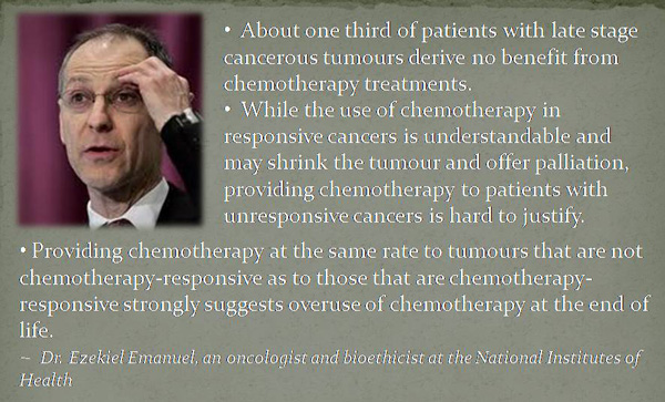
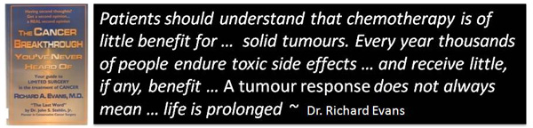
Reflect on these quotations


You must be logged in to post a comment.