Helping a Six-Year-Old with Cancer of the Brain Stem

Ros (S-321) is 6-years old. For about a year, when she was 5 years old, she had been vomiting and having headaches. In December 2012, her parent brought her to the hospital where a CT scan was performed. The doctor in Aceh, Indonesia said there was a tumour in her brain. It was not operable. Ros was asked to go to Jakarta for further treatment.

Her parent decided to bring her to Kuala Lumpur instead.  MRI performed on 14 December 2012 indicated a well-defined mass measuring 3.2 x 3.5 x 2.9 cm, compressing onto the adjacent 4th ventricle. The lateral and 3rd ventricles were dilated. This was diagnosed as pontine tumour.

Composite

The doctor said the tumour was not operable and there was also no medication for her. Back in Aceh, Ros’s parent started her  on herbs like Soursop leave tea, Sarang Semut, etc.

On 20 January 2013, her father came to Penang to seek our help. Surprisingly, Ros did not have much symptoms. She would only suffer headaches if she had fevers. She had squint or cock eyes, otherwise she was alright.


Ros was prescribed Capsule A, Brain Tea 1 and 2 plus C-tea and Brain Leaf. Due to her age, she was asked to take only at half dosage. We warned her father that the herbs are bitter and his daughter might have difficulty taking them. His reaction was, We will try.


Our Herbs Helped Her

To our surprise Ros’s auntie came back to see us on 22 February 2013 – a month after taking the herbs. Ros did not have difficulty drinking the bitter and awful tasting herbal teas! And she had improved. There were no more fevers. Her eye movements had improved. There no more headaches or vomiting. Indeed the herbs had helped Ros tremendously.

There was one problem though! The family wanted Ros to bring fried bananas to school. Our answer was, Absolutely no! No fried stuff.  Since taking our herbs, Ros seemed to have frequent urination at night. Sometimes she even urinated in bed without knowing. Since Ros did not come to see us, there was nothing we could do to assess her bladder energy using the AcuGraph. We suggested that we would let the problem take care of itself.

Two months later, i.e. 26 April 2013 (and Ros was already 3 months on our herbs), Ros’s father came to see us again. He was happy and said that Ros had improved tremendously. Her eyes seemed to be normal now. If she reads too much then her eyes become juling (cockeye or squint). Otherwise she is okay. Since taking the herbs Ros did not have any more headaches. She did not vomit either.

The family had solved the problem of her night urination! If Ros were to take the herbal teas way pass 6 p.m. she would urinate frequently at night. If she was to take all teas before 6 p.m. there would be no urination problem!


Current Medical Knowledge About Pontine Cancer and Its Treatment

The brain stem consists of the midbrain, pons and medulla as shown in the diagram below.

brain pons

About Pontine Glioma

  • Pontine gliomas are cancerous tumours that originate from the part of the brain known as the pons or the brain stem. It is often referred to as diffuse pontine glioma.
  • This cancer accounts for 10 to 15 percent of all childhood brain tumours. They rarely occur in adults.
  • It affected children aged 5 to 10 years old but it can occur at any age in childhood.
What is the cause of this cancer?

Currently there is no answer!

Prognosis

  • It is a highly aggressive and difficult to treat brain tumor.
    • Prognosis is poor.
    • Survival past 12 to 14 months is uncommon.

Symptoms

Each child may experience symptoms differently.

Pontine tumors affect the cranial nerves, causing symptoms related to the nerves that supply the muscles of the eye and face, and muscles involved in swallowing. This may give rise to symptoms such as:

  • double vision,
  • squints  ( a condition where the eyes point in different directions. One eye may turn inwards, outwards, upwards or downwards while the other eye looks forward.
  • inability to close the eyelids completely,
  • dropping one side of the face,
  • facial weakness,
  • problems chewing and swallowing.

The tumor also affects the “long tracks” of the brain, with resultant

  • weakness of the arms or legs and difficulty with speech and walking.
  • weakness in the arms and legs
  • problems with walking and coordination
  • difficulty with tasks like handwriting
  • changes in personality and behaviour.

Tumors may also block the flow of cerebrospinal fluid in the brain, causing increased pressure with headaches and vomiting resulting in:

  • headaches,
  • nausea and vomiting.

Symptoms usually worsen rapidly because the tumor is rapidly growing.

Treatments options
  • Radiation therapy. Radiation therapy has been the main treatment approach.  The patients’ symptoms often improve dramatically during or after six weeks of irradiation. Unfortunately, problems usually recur after six to nine months, and progress rapidly.
  • Experimental chemotherapy.
  • Surgery is not generally possible because these tumors are widely spread within the brain stem and cannot be removed. Surgery may be possible in the few patients where the tumor is very localized. Surgical resection is not an option because of where the tumor is located. Surgery in this part of the brain can cause severe neurological damage.

According to from the Dana-Faber Cancer Institute, Boston, USA,  website many specialized brain tumor treatment centers have now specialists who deliver complementary or alternative medicines. These treatments, which may help control pain and side effects of therapy include the following.

  • acupuncture/acupressure
  • therapeutic touch
  • massage
  • herbs
  • dietary recommendations.

The above information is obtained from the following websites:

  1. Diagram from: http://www.interactive-biology.com/107/what-parts-of-the-brain-control-respiration/
  2. The Dana-Faber Cancer Institute, Boston, USA. http://www.dana-farber.org/Health-Library/Childhood-Diffuse-Pontine-Glioma.aspx
  3. The Royal Marsden, London, UK. http://www.royalmarsden.nhs.uk/cancer-information/children/pages/pontine-glioma.aspx
  4. St. Jude Children’s Research Hospital, Memphis, TN, USA. http://www.stjude.org/stjude/v/index.jsp?vgnextoid=b86c061585f70110VgnVCM1000001e0215acRCRD

Articles from the Internet: How Safe Or Unsafe Are Medical Imaging Procedures?

Yeong Sek Yee & Khadijah Shaari 

To understand more about the radiation risks from medical imaging, we recommend that you read the following articles posted in the Internet.  Just Google topics like the dangers of medical/diagnostic tests, etc, etc. There are plenty of materials to read. Here are some examples.

1.       Medical Radiation Soars, With Risks Often Overlooked

Radiation, like alcohol, is a double-edged sword. Radiation can reveal hidden problems, from broken bones and lung lesions to heart defects and tumors. But it also has a potentially serious medical downside: the ability to damage DNA and, 10 to 20 years later, to cause cancer. CT scans alone, which deliver 100 to 500 times the radiation associated with an ordinary X-ray and now provide three-fourths of Americans’ radiation exposure, are believed to account for 1.5 percent of all cancers that occur in the United States.

Although the cancer-causing effects of radiation are cumulative, no one keeps track of how much radiation patients have already been exposed to when a new imaging exam is ordered. Even when patients are asked about earlier exams, the goal is nearly always to compare new findings with old ones, not to estimate the risks of additional radiation.

Read more:  http://well.blogs.nytimes.com/2012/08/20/medical-radiation-soars-with-risks-often-overlooked/?utm_source=twitterfeed&utm_medium=twitter

2.       Radiation Risks from Medical Imaging

The FDA has put forward its plan to reduce unnecessary radiation exposure from CT scans, nuclear medicine studies, and fluoroscopy.

An individual’s chance of getting cancer from a single scan is small. But because the scans are so widely used, they cause a considerable amount of harm. One study estimated that the CT scans performed in 2007 are related to some 29,000 future cancers.

What are these tests? What are their risks? When do the tests’ benefits outweigh their risks? Here are WebMD’s answers to these and other questions.

How much radiation does a person get from medical imaging studies?

  • Getting a CT scan gives a patient as much radiation as 100 to 800 chest X-rays.
  • Getting a nuclear medicine study exposes a patient to as much radiation as 10 to 2,050 chest X-rays.
  • Getting a fluoroscopic procedure exposes a patient to as much radiation as 250 to 3,500 chest X-rays.

Moreover, doctors may prescribe scans that aren’t medically justified. And since risk from radiation exposure accumulates over a lifetime, certain scans may not be appropriate for people who’ve already had a lot of scans.

Read more: http://www.medicinenet.com/script/main/art.asp?articlekey=114953

3.       Dangers of Medical Imaging Tests and Procedures

Exposure to medical imaging radiation is a concern in both adults and children. However, radiation exposure in children is of a greater concern because they are more sensitive to radiation than adults. In addition, children have longer life expectancy than adults. With repeated exposure or accumulated exposure to radiation, children may be more likely to develop health problems in the future.

Life time risk of developing cancer increases when a patient undergoes more frequent X-ray exams and at larger doses, according to the FDA. Women who are exposed to the radiation may have higher lifetime risk for developing radiation-associated cancer than men after receiving the same exposures at the same ages.

While experts believe that the risk of developing cancer with radiation exposure is relatively small, radiation exposure through these medical imaging tests should never be taken lightly.

Read more:  http://voices.yahoo.com/dangers-medical-imaging-tests-procedures-5452681.html?cat=5

4.       A Closer Look: The Downside of Diagnostic Imaging

CT and nuclear medicine tests do have a downside, however: they deliver doses of ionizing radiation from 50 to over 500 times that of a standard x-ray, such as a chest x-ray or mammogram. Scientists have raised concerns that such large doses of radiation plus the widespread and increasing use these diagnostic procedures may, in a small but significant way, pose a cancer risk in the general population.

“The use of CT in particular has gone up dramatically, and we’ve drastically lowered the threshold for using it,” said Dr. Rebecca Smith-Bindman, a visiting research scientist with NCI’s Radiation Epidemiology Branch (REB). “There’s a general belief that if you get a CT scan, you must be reasonably sick and must really need it. This is no longer true, and we are increasingly using CT scans in patients who are not that sick. There’s been drift not only in how often we use it but in how we use it.”

“We’ve only talked about the benefits of CT for the past 20 years, without considering any potential harm” she continued.

Research estimated that approximately 29,000 future cancers could be related to CT scans performed in the United States in that year alone, with women being at higher risk than men. About 35 percent of these cancers were projected to be related to scans performed in patients 35 to 54 years old, and 15 percent related to scans performed in children younger than 18. 

The medical community has proposed many ways to reduce radiation exposure from diagnostic medicine without negatively impacting the quality of patient care:

  • Reduce the number of CT exams by using other technologies (such as ultrasound or MRI) in cases where they would provide equal diagnostic quality.
  • Limit the use of CT in healthy patients who would obtain little benefit (such as whole-body CT screening).
  • Limit the use of repeat CT surveillance of patients in whom a diagnosis has already been made, when repeat scanning would lead to little change in their treatment.
  • Track and collect information on radiation exposure for individual patients

Read more: http://www.cancer.gov/aboutnci/ncicancerbulletin/archive/2010/012610/page8

5.       Ionizing Radiation Exposure with Medical Imaging

Medical diagnostic procedures used to define and diagnose medical conditions are currently the greatest manmade source of ionizing radiation exposure to the general population. The risks and benefits of radiation exposure due to medical imaging and other sources must be clearly defined for clinicians and their patients.

Radiation damages the cell by damaging DNA molecules directly through ionizing effects on DNA molecules or indirectly through free radical formation. A lower dose delivered through a long period of time theoretically allows the body the opportunity to repair itself. Radiation damage may not cause any outward signs of injury in the short term; effects may appear much later in life.

Medical ionizing radiation has great benefits and should not be feared, especially in urgent situations. Obviously, using the lowest possible dose is desired. In fact, a central principle in radiation protection is “as low as reasonably achievable.” Therefore, the prescribing physician must justify the examination and determine relevant clinical information before referring the patient to a radiologist. Indications and decisions should reflect the possibility of using non-ionizing radiation examinations, such as MRI or ultrasonography.

Repetition of examinations should be avoided at other clinics or sites.

The International Commission on Radiological Protection (ICRP) estimates that the average person has an approximately 4-5% increased relative risk of fatal cancer after a whole-body dose of 1 Sv.

X-rays (including CT scans) should be ordered judiciously. An article in the New England Journal of Medicine notes that the evidence is “convincing” that the radiation dose from CT scans can lead to cancer induction in adults and “very convincing” in the case of children. Clinicians need to realize that doses from a typical CT scan can range from 6-35 times higher than the dose of a standard chest x-ray examination.

Read more:  http://emedicine.medscape.com/article/1464228-overview#a30

6.  Doctors Order More Tests when They Benefit Financially: Ask If You Really Need that Test Your Doctor Ordered

Researchers from the Institute for Technology Assessment at the Massachusetts General Hospital Department of Radiology found that there was no mistaking that diagnostic imaging tests were being ordered far more than they deemed necessary. The question that begs to be answered is, “why?”

Many doctors referred their patients to imaging centers that were affiliated with their practice, or were even done by the doctor’s own staff. When a physician has such a close relationship with the provider conducting the imaging study, there is the possibility that the physician will benefit financially from ordering additional imaging studies.

Read more: http://voices.yahoo.com/doctors-order-more-tests-they-benefit-financially-631960.html?cat=5

7.       Radiation Danger from CT and PET Scans

A recent study in the New England Journal of Medicine has found a significant link between radiation exposure and imaging procedures such as CT and PET scans. The use of such technologies has grown from just 3 million in 1980 to 67 million in 2006, and has contributed, some estimate, to upwards of 2% of fatal cancer cases.

Studies have shown that there is little consumer understanding of the risks involved in being subject to such procedures.

Dr. Harlan M. Krumholz proffers that the use of CT scans is increasing because they have become part of our culture. “People use imaging instead of examining a patient; they use imaging instead of talking to the patient,” (New York Times, Study Finds Radiation Risk for Patients, August 27, 2009). For these reasons, imaging technologies have become a common diagnostic tool even when they are not required.

Read more: http://blog.hcfama.org/2009/08/27/radiation-danger-from-ct-and-pet-scans/

8.       Study Finds Radiation Risk for Patients

At least four million Americans under age 65 are exposed to high doses of radiation each year from medical imaging tests, according toa new study in The New England Journal of Medicine. About 400,000 of those patients receive very high doses, more than the maximum annual exposure allowed for nuclear power plant employees or anyone else who works with radioactive material.

Dr. Rita Redberg, a cardiologist and researcher at the University of California, San Francisco, who has extensively studied the use of medical imaging, said it would probably result in tens of thousands of additional cancers. It’s certain that there are increased rates of cancer at low levels of radiation, and as you increase the levels of radiation, you increase cancer.

Dr. Reza Fazel, a cardiologist at Emory University, said the use of scans appeared to have increased even from 2005 to 2007, the period covered by the paper. “These procedures have a cost, not just in terms of dollars, but in terms of radiation risk.”

Read more: http://www.nytimes.com/2009/08/27/health/research/27scan.html?_r=0

9.      Radiation Exposure from Medical Diagnostic Imaging Procedures

Ionizing radiation is used daily in hospitals and clinics to perform diagnostic imaging procedures.

Which types of diagnostic imaging procedures use radiation?

•  In x-ray procedures, x rays pass through the body to form pictures on film or on a computer or  television monitor, which are viewed by a radiologist. If you have an x-ray test, it will be performed with a standard x-ray machine or with a more sophisticated x-ray machine called a CT or CAT scan machine.

• In nuclear medicine procedures, a very small amount of radioactive material is inhaled, injected, or swallowed by the patient. If you have a nuclear medicine exam, a special camera will be used to detect energy given off by the radioactive material in your body and form a picture of your organs and their function on a computer monitor. A nuclear medicine physician views these pictures. The radioactive material typically disappears from your body within a few hours or days.

Do magnetic resonance imaging (MRI) and ultrasound use radiation?

MRI and ultrasound procedures do not use ionizing radiation. If you have either of these types of studies, you are not exposed to radiation.

There is no conclusive evidence of radiation causing harm at the levels patients receive from diagnostic xray exams. Although high doses of radiation are linked to an increased risk of cancer, the effects of the low doses of radiation used in diagnostic imaging are not known.

Read more: https://hps.org/documents/meddiagimaging.pdf

10.        Radiation Risk of Medical Imaging for Adults and Children

Which kinds of tests are associated with Ionising radiation and which ones are not?

1.  X-rays
X-rays are ionising radiation produced by equipment used in the following types of procedures:

  • Computed tomography (CT)
  • Fluoroscopy (where the image produced by the X-ray beam is made into a moving picture on a TV screen
  • Plain radiology/X-ray film, digital and computed radiography (see  Plain Radiography / X-rays)
  • Mammography (see Diagnostic Mammography)
    • The radiation exposure from having an X-ray, fluoroscopy, mammography or CT examination only occurs while the machine is on.

2.  Magnetic resonance imaging (MRI)
MRI uses strong magnetic fields and radio waves to produce images. It does not use ionising radiation (see Magnetic Resonance Imaging (MRI)).

3.  Ultrasound
Ultrasound uses high frequency sound waves that the human ear cannot detect to obtain imaging information (see Ultrasound).

4.  Nuclear medicine
Nuclear medicine is a medical specialty that involves the administration of a small amount of a radioactive material into the patient. The patient becomes weakly radioactive for a short time and images are made from the radiation given off from the patient (see Nuclear Medicine).

How do I decide whether the risks are outweighed by the benefits of exposure to X-radiation when I have a radiology test or procedure?

  • Ask your doctor about the procedure and how it will help to provide information about your symptom or the presence of disease or injury.
  • Ask your doctor about the risks of the procedure and what the risks would be of not having the procedure, i.e. if your doctor needs the information in order to identify and plan the most appropriate treatment.

While there is a small risk of harm from ionising radiation, there could be a greater risk of not having the information, e.g. failure to detect potentially serious disease that may be easily treated at an early stage but harder to treat or incurable if detected later.

It may also be as beneficial to you to confirm the absence of disease or injury as it is to confirm its diagnosis.

Read more: http://www.insideradiology.com.au/pages/view.php?T_id=57

11.   Radiation in Medical Imaging Has Its Risks

Almost all medical procedures, including imaging procedures that use radiation, have risks associated with them. Physicians and patients should carefully consider the potential benefits and the risks when considering the use of imaging techniques that involve radiation.

Here are some things for healthcare providers to consider when deciding whether or not an imaging procedure that uses medical radiation is the right choice.

  • What is the purpose of the procedure? For example, is it to arrive at a diagnosis, assess treatment response, or is it preventive screening?
  • Are there alternative imaging procedures that could accomplish the same goal without medical radiation, such as ultrasound or magnetic resonance imaging?
  • What are the risks of not having the imaging procedure done?
  • How old is the patient? The risks for pediatric and adolescent patients may be different than for adults.
  • Is the patient pregnant, possibly pregnant, or breastfeeding?
  • What other procedures is the patient likely to undergo during this workup?
  • What is this person’s radiation exposure from previous medical procedures? For example, has the person undergone multiple CT or nuclear medicine scans in the past?
  • What is this person’s occupational exposure to radiation, if any?
  • Will the imaging exam be performed on low-dose equipment?

The standard unit of measure for radiation absorbed by an individual is called the “Sievert,” or Sv (sometimes identified by a smaller unit called the “millisievert,” or mSv). Common medical imaging tests such as X-rays or mammograms generally expose patients to a radiation dose of less than 1 mSv.

Other procedures using CT, nuclear stress tests, or fluoroscopy-guided exams often involve radiation in the range of 5-40 mSv.

single exposure at these diagnostic levels may not pose much risk to the patient. But when a patient has numerous tests over a period of time, the cumulative exposure may raise the level of risk. To minimize cumulative exposure, physicians should determine whether a procedure using medical radiation is necessary to achieve the diagnosis or whether an alternative imaging procedure may offer the same diagnostic benefit.

Read more:  http://www.gehealthcare.com/dose/medical-radiation/benefits-and-risks.html

12.   How Safe or Unsafe Are Medical Imaging Procedures?

Radiation exposure is a known risk factor for cancer. Recent estimates suggest, for example, that as many as two percent of cancers could be attributed to radiation during CT scans. Although the radiation exposure from a single test is minimal, the frequency of the use of imaging tests that emit radiation continues to grow expansively, and often patients undergo repeated or multiple types of tests, thereby increasing their cumulative exposure to potentially cancer-causing radiation.

Read more:  http://www.sciencedaily.com/releases/2009/08/090826191837.htm

Advice to Patients

Lately, we have noticed that certain medical centres have been urging   cancer patients to perform regular CT or PET scans (some every 3 months)  to “monitor” the progress of their cancer treatment. Sometimes some cancer patients think that such CT/PET scans are “treatment” itself. The medical establishment obviously have a financial benefit in urging you to perform more imaging/diagnostic procedures.

When deciding whether or not to perform further imaging/diagnostic procedures, we would advise you to seek answers to the following:

  • What is the purpose of the procedure? For example, is it to arrive at a diagnosis, assess treatment response, or is it preventive screening?
  • Are there alternative imaging procedures that could accomplish the same goal without medical radiation, such as ultrasound or magnetic resonance imaging?
  • What are the risks of not having the imaging procedure done?
  • What is your radiation exposure from previous medical procedures? For example, have you undergone multiple CT or nuclear medicine scans in the past?

Each time you are asked to do a CT Scan/PET Scan, be aware of the amount of radiation that would be bombarding your body and do remember that the radiation is accumulative i.e. it accumulates in your body, not the doctor’s body (except his wallet gets heavier). The following article is self explanatory:

Video Presentation: Talk by Chris Teo, 11 May 2013 Kuala Lumpur

1 Title

Pt 1 Get The Best Of Both Worlds

Pt 2 Die of Cancer or Of Treatment

Pt 3 Overblown Statistics and Empty Promise

Pt 4 Doc, Give Me An Honest Answer

Pt 5 Beware Expensive and Dangerous Drug That Does Not Cure

Pt 6 Does Chemo Make Sense? Are Doctors Truly Honest?

Pt 7 Chemo Treats or Promotes Cancer?

Pt 8 Don’t Panic, Heal Yourself

Pt 9 Believe the Diagnosis Not the Prognosis

Pt 10 Chemo Almost Kill, Herbs Kept Him Alive

Pt 11 To Live or Die Is Your Choice

Pt 12 Recovered:  Even After Doctor Said No Chance

Pt 13 Doctor’s Bullying Ways and Self-interest

Pt 14 The CA Care Therapy

Chemotherapy Spreads Cancer and Makes It More Aggressive: Articles From the Internet

Compiled by Yeong Sek Yee & Khadijah Shaari

1.   Perilous Approach: Avastin and Sutent Promote Growth of Breast Cancer Stem Cells

(Note: Many patients in Malaysia have been treated with this very expensive drug)

The U.S. Food and Drug Administration recently revoked approval of Avastin for treating breast cancer. The reversal was in response to clinical trials showing that the drug’s benefit was short-lived, with breast cancer patients quickly relapsing and the cancer becoming more invasive and metastatic.

Cancer treatments designed to block the growth of blood vessels were found to increase the number of cancer stem cells in breast tumors in mice, suggesting a possible explanation for why these drugs don’t lead to longer survival, according to a study by researchers at the University of Michigan Comprehensive Cancer Center.

While anti-angiogenic drugs do shrink tumors and slow the time until the cancer progresses, the effect does not last, and the cancer eventually regrows and spreads.

The researchers treated mice with breast cancer using the anti-angiogenesis drugs Avastin (bevacizumab) and Sutent (sunitinib). The researchers found that tumors treated with these drugs developed more cancer stem cells, which fuel a cancer’s growth and spread and are often resistant to standard treatment.

Read more: http://www.med.umich.edu/cic/2012-spring/perilous-approach.html

2)      Chemotherapy Can Make Cancers More Resistant To Treatment And Even Encourage Them To Grow

  • Chemotherapy treatment for some cancers may actually encourage tumours to grow, researchers have claimed.
  • The treatment triggers the healthy body cells around the tumour to produce a protein that helps the disease to resist treatment.
  • The surprise discovery suggests that some forms of the cancer treatment are doing more harm than good. 

Read more:  http://www.dailymail.co.uk/health/article-2184277/Chemotherapy-encourage-cancer-growth.html 

3)      Chemo Does Not Cure: Often It Inflicts Damage and Spreads Cancer

  • German investigators from Friedrich-Schiller University in Jena, have shown that Taxol (the “gold standard of chemo”) causes a massive release of cancer cells into circulation.
  • Such a release of cancer cells would result in extensive metastasis months or even years later, long after the chemo would be suspected as the cause of the spread of the cancer. This little known horror of conventional cancer treatment needs to be spread far and wide, but it is not even listed in the side effects of Taxol.

 Read more:  http://www.tbyil.com/Chemo_Does_Not_Cure.htm

4)      Chemo/Radiation “Therapy” May Fuel Cancer Spread

  • Treatment may fuel cancer’s spread
  • Treating cancer with surgery, chemotherapy or radiation may sometimes cause tumors to spread, researchers say.
  • Tests in mice show that using the chemotherapy drug Doxorubicin or radiation both raised levels of TGF-beta, which in turn helped breast cancer tumors spread to the lung.

Read more: http://rense.com/general76/fuel.htm

5)      Chemotherapy Causes Resistance and Spread of Cancer

Peter Nelson of the Fred Hutchinson Cancer Research Center in Seattle co-authored a study and published it in Nature Medicine this month detailing how chemotherapy not only produces resistance to chemotherapy by cancerous tumors but also stimulates its growth and metastasis (spread).  Approximately 90% of people with metastatic cancer become resistant to chemotherapy.  This occurs readily in cancers of the breast, prostate, lung, pancreas and colon.

Read more: http://www.anoasisofhealing.com/the-chemotherapy-cover-up/#axzz2S15t3mvT

6)      Chemo Could Spread Cancer.

new research published in Nature Medicine shows that chemotherapy can actually be extremely counterproductive in treating cancer as it could spur healthy cells to release a compound that actually stimulates cancer growth.

Read more: http://personalliberty.com/2012/08/07/chemo-could-spread-cancer/

7)      Chemotherapy can Backfire and Encourage Cancer Growth

Chemotherapy can backfire by triggering healthy cells to secrete a protein that sustains tumor growth, which could explain why some patients become resistant, a new study suggests.

Read more: http://www.medicaldaily.com/articles/11314/20120806/cancer-chemotherapy-resistance-immunity-nature.htm#BVXeMw9SBDTZpR0W.99

8)      Study Links Cancer’s Ability to Spread with Chemotherapy Resistance

Doctors who treat patients with breast cancer have known that tumors that develop resistance to chemotherapy are also more likely to grow larger and to spread, or metastasize, to other parts of the body.

Read more: http://www.mskcc.org/blog/study-links-s-ability-spread-chemotherapy-resistance

9)       Anti-Cancer Drugs Make Tumors More Deadly

…..new research shows that aggressive treatment (used to shrink or remove even relatively small, slow-growing or encapsulated, harmless tumors) may create a situation where the entire body is riddled with highly aggressive cancers.

This study, published in the January 17, 2012 issue of Cancer Cell,finds that a group of little-explored cells that are part of every primary cancerous tumor likely serve as important gatekeepers against cancer progression and metastasis.

Read more: http://www.ener-chi.com/anti-cancer-drugs-make-tumors-more-deadly/

10)   Anti-Cancer Drugs Make Tumors More Deadly

  • Just imagine you were diagnosed with a cancerous tumor, and your doctor told you that his/her proposed treatment could reduce the size of your tumor by 30 percent, but at the same time increase your chances of developing secondary tumors by a whopping 300 percent!
  • That is exactly what is demonstrated in recent research (at Harvard and MD Anderson Cancer Centers), and published in conventional Oncology Journals! The history of conventional anti-cancer therapies is replete with cases where the treatment turned out to be far more devastating than the disease itself.

Read more: http://hbmag.com/anti-cancer-drugs-make-tumors-more-deadly/

11)   Exposed: Deadly Cancer Drugs Make Cancer Worse and Kill PatientsMore Quickly

  • Cancer drugs, pushed by many drug companies as the only ‘scientific’ method of combating cancer alongside chemotherapy, have been found to actually make cancer worse and kill patients more quickly.
  • The findings come after research was conducted on the cancer drugs at the Beth Israel Deaconess Medical Center in Boston. Sold at a premium price to cancer sufferers, it turns out these drugs are not only ineffective but highly dangerous.

Read more: http://naturalsociety.com/deadly-cancer-drugs-make-cancer-worse-and-kill-patients-more-quickly/

12)   Breaking News: Cancer Drugs Make Tumors More Aggressive And Deadly

…….scientists at the University of Alabama at Birmingham (UAB) Comprehensive Cancer Center and UAB Department of Chemistry are currently investigating the very real possibility that dead cancer cells left over after chemotherapy spark cancer to spread to other parts of the body (metastasis).

·         A study just published in the January 17 issue of the journal Cancer Cell concludes that anti-angiogenic therapies (which shrink cancer by cutting off tumors’ blood supply) may be killing the body’s natural defense against cancer by destroying pericyte cells that likely serve as important gatekeepers against cancer progression and metastasis.

Read more:  http://www.infowars.com/breaking-news-cancer-drugs-make-tumors-more-aggressive-and-deadly/

13)   Woops! Study Accidentally Finds Chemotherapy Makes Cancer Far Worse.


  • A team of researchers looking into why cancer cells are so resilient accidentally stumbled upon a far more important discovery.

While conducting their research, the team discovered that chemotherapy actually heavily damages healthy cells and subsequently triggers them to release a protein that sustains and fuels tumor growth. Beyond that, it even makes the tumor highly resistant to future treatment.

Read more:  http://naturalsociety.com/chemotherapy-makes-cancer-far-worse/

14)   Chemotherapy Backfires – Causes Healthy Cells To Feed Growth Of Cancer Tumors

 This protein,dubbed “WNT16B,” is taken up by nearby cancer cells, causing them to “grow, invade, and importantly, resist subsequent therapy,” said Peter Nelson of the Fred Hutchinson Cancer Research Center in Seattle. He’s the co-author of the study that documented this phenomenon, published in Nature Medicine. This protein, it turns out, explains why cancer tumors grow more aggressively following chemotherapy treatments. In essence, chemotherapy turns healthy cells into WNT16B factories which churn out this “activator” chemical that accelerates cancer tumor growth.

Read more:  http://www.naturalnews.com/z036725_chemotherapy_cancer_tumors_backfires.html

15)          Chemo and Radiation Actually Make Cancer More Malignant

The very treatments may have transformed a relatively slow growing tumor into a rapidly proliferating and invasive one.

Read more: http://www.greenmedinfo.com/blog/chemo-and-radiation-actually-make-cancer-more-malignant

16)         Beating Cancer With Chemotherapy And Better Drugs: Junk Science?

  (1) The Department of Oncology at North Sydney Cancer Centre in 2004 published a report evaluating chemotherapy over the years and concluded that ‘it only made a minor contribution to survival’. The figures they came up with were 2.3% in Australia and 2.1% in America.

(2) The Fred Hutchinson Cancer Center in Seattle that concluded ‘Chemotherapy can cause cancer to return’. They said CAUSE – apparently chemotherapy can cause healthy cells to produce a protein WNT16B and this is taken up by cancer cells – it helps them re-grow and even protects them from the next round of chemotherapy.

(3)  A ‘landmark study’ from their Cambridge Institute showed there were 10 different ‘clusters’ of breast cancer types. ‘No longer does one size fit all’ they cried.

 (4) Three research studies reported on the existence of Cancer Stem Cells at the heart of tumours. A couple of UK cancer centres (Bart’s Hospital and the Blizzard Institute, London) have even isolated these nasty little cells. Apparently, if you don’t kill them off, they can re-grow. In one of the three studies (from the University of Texas South Western Medical Centre), there were statements such as ‘Cancer Stem cells are in charge of tumours’, and the lead researcher, Dr Louis Parada and the other researchers added, ‘In the past we have tried to get rid of the entire stew of cancer cells. But shrinking a tumour by 50% is irrelevant. No current drugs tackle cancer stem cells.

 (5)  Perhaps the final words should therefore go to Duke’s University Medical Centre in Carolina who in their 2012 report concluded that ‘Patients with cancer are largely being mislead into believing that the drug they are being offered is somehow going to cure them’.

Read more: http://www.junkscience.co.uk/2013/04/junk-science-number-50-beating-cancer-with-chemotherapy-and-better-drugs/

Lung-Liver Cancer: When Everything Went Wrong for Her

KF (S-364) is a 40-year old Indonesian lady. In 2011, she had coughs for more than a year. There was no blood in her phlegm. She took cough syrup but was not effective. In January 2013, she became breathless and found it difficult to climb the stairs. Her problem became more serious and she went to a private hospital in Tangerang. There was fluid in her lungs. Pleural tapping was performed once. But this procedure did not help her much. She was still breathless. Another tapping was done but there was no fluid.

A CT scan was performed and the doctor said there was a tumour. She was referred to a lung surgeon who told her that surgery was not indicated because her lungs could be filled with fluid and there was “not enough preparation” for him to proceed with the surgery.

Not satisfied, KF went to another lung specialist in a Jakarta hospital. Another CT scan was done, specimens were collected, etc., but the specialist could not determine the cause of her problem. The lung specialist suggested that KF’s problem could be due to “jamur” or parasites! KF was prescribed antibiotics. KF was also asked to do a biopsy, which she declined.

KF came to a private hospital in Penang in April 2013. She consulted a lung specialist. A CT done on 5 April 2013 showed:

  • Extensive circumferential heterogeneously enhanced lobulated masse in the left hemithorax.
  • There is a central fluid / necrotic area seen
  • Compression of the left hilar vessels and bronchi
  • There is infiltration into mediastinum
  • Trachea, oesophagus and heart are displaced to the right side
  • Suspicious left pericardial invasion seen
  • A faint hypodense nodule seen in segment 8 of the liver measuring 15 mm in diameter.
  • Impression:  Large circumferential left hemithorax mass with liver metastasis and suspicious left pericardial invasion. Differential diagnosis: 1. Mesothelioma  2. Bronchogenic carcinoma. 

s364-a

s364-b

A biopsy of the left chest tumour indicated poorly differentiated adenocarcinoma infiltrating the chest wall.

Immunohistology report of 20 April 2013 indicated the cells are positive for CK7 only and negative for CK20 and TTF-1.

The lung specialist referred her to a surgeon who told her surgery was not indicated for her case. KF was then referred to an oncologist. KF was prescribed 5 type of medication and one of which was Iressa. KF was asked to go home and try the Iressa and see what happen. If Iressa was not effective, KF would have to undergo chemotherapy.

A week in the private hospital cost RM 21,000. In addition she paid RM 7,000 for the medication inclusive of a month’s supply of Iressa.

KF was told Iressa would cause side effects. And she was not willing to take it. Someone living in the same apartment as she, told her about CA Care. She came to seek our help on 25 April 2013. She presented with the following:

  • Difficulty sleeping
  • No appetite
  • Tiredness and lack of energy
  • Difficulty breathing
  • Cough throughout the night with white phlegm.

We prescribed KF Capsule A, B, C, D and E. In addition she has to take many teas: Lung 1 and 2, Lung Phlegm, Liver 1 and 2. She was given Cough 5 for her coughs (white phlegm).

What had gone wrong?

  1. She went to the hospital in Tangerang. Fluid was tapped out but she did not improve much. The doctor could not say if it was cancer or not. Not satisfied she went to another hospital in Jakarta. Here again there was not much help. One doctor even suggested that the problem could be due to “jamur” or parasites. I wonder how the lung expert could ever give such a suggestion! Anyway the anti-jamur medication did not work for her. 
  2. The patient came to a hospital in Penang. After a week stay she was discharged and was still not satisfied. She came to CA Care for help.
  3. A week’s stay in the hospital cost her RM 21,000. Did she get any better? The lung specialist could not help. She was referred to a surgeon who could not solve her problem either. The next obvious stop was the oncologist. The oncologist offered five medications one of which was Iressa. The total cost of the medication was RM 7,000. She was told to try out Iressa for a month and see if this could help her!  But she was not keen on Iressa because of the possible side effects. We felt sorry for KF – having made to pay for such an expensive medication which she was not willing to take.
  4. Was Iressa prescribed based on “scientific” fact or on a trial and error basis? Immunohistology showed that cells were only positive for CK7 and negative for CK20 and TTF-1. Is this the kind of cancer that would respond to Iressa?  I also do get patients who told me that she/he was asked to take Iressa in spite of the fact that test showed that the cells were negative for Iressa. But the justification given was that even for such “negative” cases Iressa seemed to work on some patients. Looks like we have to throw science out of the window!

Notes on The Epidermal Growth Factor Receptor (EGFR)

According to Kakiuchi et all, Gefitinib (Iressa), has shown potent anti-tumor effects and improved symptoms and quality-of-life of a subset of patients with advanced non-small cell lung cancer (NSCLC). However, a large portion of the patients showed no effect to this agent.  http://www.ncbi.nlm.nih.gov/pubmed/15496427

Lung adenocarcinomas with mutated epidermal growth factor receptor have significant responses to tyrosine kinase inhibitors, although for unselected patients it does not appear to have a survival benefit. Both EGFR mutation and gene amplification status may be important in determining which tumors will respond to tyrosine kinase inhibitors. http://www.jthoracdis.com/article/view/87/152

The tumors that responded to the EGFR TK inhibitors (TKIs) gefitinib and erlotinib contain somatic mutations in the EGFR TK domain. The two most common EGFR mutations are short in-frame deletions of exon 19 and a point mutation (CTG to CGG) in exon 21 at nucleotide 2573. Together, these two types of mutations account for ~90% of all EGFR mutations in NSCLC. Other recurrent but far less common EGFR mutations known to be associated with sensitivity to EGFR TKIs include mutations in exon 18 and in exon 21. Screening for common EGFR mutations in patients with lung adenocarcinomas can now be performed in clinical molecular diagnostic laboratories to predict which patients will respond to EGFR TKIs. It can be performed on archival material as well as on fine-needle biopsies. http://www.nature.com/modpathol/journal/v21/n2s/full/3801018a.html

Predicting Sensitivity to Iressa and Tarceva

Iressa (gefitinib) and Tarceva (erlotinib) were being tested in large numbers of patients with advanced non-small cell lung cancer.  Iressa did not improve overall survival compared to placebo treatment in previously treated NSCLC  patients.

However, about 10% of Western patients treated with either of these drugs had dramatic and sometimes long-lasting responses. Investigators at the Dana Farber Cancer Institute, Massachusetts General Hospital in  Boston, and also at Memorial Sloan Kettering Cancer Center in NYC published results showing that most of these “dramatic responders” had recurring mutations in the tyrosine kinase (TK) domain of the EGFR gene.

In the NSCLC patients who have mutations in the TK domain of the EGFR. This makes the cancer cell exquisitely sensitive to dying when the switch is turned off by a drug like Iressa or Tarceva, and explains why some patients can do so well on these drugs. Although there can be mutations anywhere in the TK domain, only some of them confer sensitivity to the TKIs.

About 45% of sensitizing mutations are what are called in frame deletions in exon 19, making them the most common EGFR mutations. About 40-45% of the sensitizing mutations are point mutations in exon 21. Most of the remaining mutations don’t cause the EGFR to be sensitive to EGFR TKIs.

A point mutation in exon 20 resulting seems to allow the EGFR TK to work much better than normal. Mutations in exon 20 have also been associated with resistance.

Mutations can be detected using sequencing to identify every mutation in the tyrosine kinase domain, whether predictive of responsiveness to TKIs or not. Another method is something called allele-specific polymerase chain reaction (PCR) which can then be detected by a machine. This method only detects 28 of the most common EGFR mutations, but generally requires smaller amounts of tissue than sequencing and has a slightly faster turnaround time. There is also evidence that this method may be more sensitive than direct sequencing.

Quoted from: http://cancergrace.org/lung/2010/10/10/overview-of-molecular-markers-in-lung-cancer/

Read more:  Practical Management of Patients With Non–Small-Cell Lung Cancer Treated With Gefitinib  http://jco.ascopubs.org/content/23/1/165.full


WHY CONVENTIONAL CANCER TREATMENT IS FATALLY FLAWED – A DOCTOR’S VIEW

By Yeong Sek Yee and Khadijah Shaari

Is conventional cancer treatment really fatally flawed? This seems to be the opinion of Dr Margaret I. Cuomo, MD who wrote the article “Why Cancer treatment is Fatally Flawed” (Copy of article attached or view article at the following link): http://www.huffingtonpost.com/margaret-i-cuomo-md/cancer-prevention_b_1609446.html

Curious to find out more, we subsequently bought the book, “A WORLD WITHOUT CANCER” by Dr Margaret Cuomo, a board – certified radiologist (more details at the end of this article).

World wiithout cancer2

We would like to summarize some of the main points of Chapter 4 of this book which is surprisingly entitled “CUT, BURN AND POISON: A LOOK AT TODAY’S TREATMENT OPTIONS.” The following points sum up Dr Cuomo’s disappointment in conventional treatment (in her own words): –

a)      For most of the 20th Century, we have used 3 basic approaches to treat cancer: surgery, chemotherapy and radiation. With experimentation and practice… some modalities may have improved, yet…our results remain entirely inadequate. In crude fashion, we are still trying to cut, poison, and burn our way through cancer (pages 56/57)

b)      One cancer expert has called our approach to cancer “damage control”. We deploy the heavy artillery to kill as much of the cancer as we can, hoping that it doesn’t escape our weaponry. However, all too often it does (page 10).

c)      When it comes to treating cancer, we seem to be in a holding pattern (page13) Cancer has turned out to be far more clever than we imagined… it is able to develop resistance even to highly toxic drugs… it has an uncanny ability to outwit the therapeutics sent to inhibit its growth (pages 12/13).

What are Dr Cuomo’s views on cut, poison, and burn (or alternatively known as surgery, chemotherapy and radiation)?

a)      Cutting : The Surgical Option          

  • Sometimes, surgery isn’t an option. Tumors may be inoperable because they are weakened in an area that’s almost impossible to reach, or a patient may not be strong enough to withstand surgery (page 64).
  • Even if someone does undergo surgery, there is no way to be sure it has been successful. Although every cancer patient yearns to hear the words “we got it all,” cancer has the capacity to work in nearby tissues, even where a clear margin seems to have been obtained, or to spread undetected through the blood or lymph nodes (pages 64/65)
  • Today’s surgery may be safer than it was, but in many cases, it still doesn’t cure the disease. Cutting out organs and tissues does little to fight cancers peripatetic capacity to travel through the blood to colonize other parts of the body (page 13).

b)      Poison: The Limits of Anti Cancer Drugs

  • Pharmaceuticals, collectively called systemic therapy “include 3 major groups : chemotherapy, targeted therapy, and hormonal therapy (page 65).
  • Whether administered as neo-adjuvant therapy (before surgery or radiation) or adjuvant (after surgery or radiation), physicians sometimes avoid the technical language and simply call them “poison” – what we want the drug to do is kill cancer cells. Unfortunately, they can kill normal cells as well… “Chemotherapy is like taking a stick and beating a dog to get rid of fleas” (page 65).
  • Chemotherapy can be especially grueling, with many patients receiving one drug after another. “Usually the first treatment regimen works and then it stops working after a certain point” says Dr Nicholas Vogelzang of the American Society of Clinical Oncology (page 57).
  • The toxic effects of chemotherapy are not limited to cancer cells. Every drug has its own package of debilitating and sometimes bizarre side effects. Some of these occur during treatment and immediately afterwards, some occur five or more years later (page 67).
  • According to the American Society of Clinical Oncology, late effects can strike just about anywhere in the body – including the heart, lungs, brain and spinal cord, bones and joints, endocrine and digestive system. Even dental and vision problems can occur (page 68).
  • When it comes to chemotherapy, no patient emerges unscathed, and the benefits may not last. If cancer cells became resistant to chemotherapy, as often happens over time, they begin to multiply again. The question for patients is always whether the gain in longevity will ultimately outweigh the brutality of the treatment (pages 69/70).
  • Even drugs used to treat the side effects of chemotherapy have been linked to secondary cancers (page 69).
  • Most of the drugs we have to fight solid tumors influence a specific part of a cellular cycle and have only a transient effect. They don’t stop the progress of a tumor indefinitely. A slow growing tumor is still growing and is perhaps beginning to spread undetected elsewhere in the body (page102).
  • Lastly, many drug therapies are not even studied on the basis of their impact on overall survival, but rather on their impact on progression-free survival or the maintenance of stable disease. There is a big difference (page 101).

c)      Burn: Radiation Therapy

  • Although we have gotten much better at aiming radiation directly at a tumor, we can’t target it with the precision necessary to burn only cancer cells and leave healthy ones intact. That means that this therapy, too, is damaging (page 75).
  • Months or even years after radiation, chronic side effects can surface. With radiation, as with chemotherapy, the list of potential problems is lengthy (page77).
  • Most troubling is the possibility that other cancers will emerge, often near the original site, long after receiving radiation therapy…children who survived cancer were 15 times more likely to die of a subsequent cancer later in life (page 79).
  • Though the young are most vulnerable, secondary cancers can develop as a result of radiation at any age.

Flawed Clinical Trial Design:

Besides cancer treatments being fatally flawed, clinical trial designs are also flawed in the first place (page138). According to Dr Cuomo:

  • Good medicine is built on clinical trials in which promising scientific discoveries are tested in human beings (page 138).
  • Too often, clinical trials fail to discover who benefit from a therapy and who will be harmed (page140)….we need to be doing the studies that will tell us which patients require chemotherapy (page 141).
  • ….because we don’t know the profile of those who will gain, almost all patients routinely get chemotherapy……that translates into giving therapies that fail 95% of the time in order to find that 3 to 5% of responsive patients (page 141).

Some other pertinent remarks of Dr Cuomo that are particularly succinct: –

  •  The fact that we have made so little progress after a century of using the same basic techniques surely suggests that we aren’t taking the right approach. The problem? “Even one cancer cell can lead to death” says James Holland, MD a distinguished professor of neoplastic diseases at Mt. Sinai School of Medicine in New York City. Relapse is always a possibility until we can guarantee that there are no cancer cells in the body (page 79).
  • When death can come from a single cell that successfully eludes our most brutal attempts to cut, poison, and burn it the sword of Damocles hangs over every patient’s head (page 59).
  • We should differentiate between drugs that make a small difference and drugs that make no difference at all. A lot of cancer drugs are not worth very much in terms of prolongation of life (page 110).
  • Meanwhile, our continuing emphasis on producing, prescribing, and paying for one marginally useful cancer therapy after another suggests that we are in the wrong playing field (page 124).

In conclusion, when Dr Cuomo commented in The Final Word (page 244) that, when President Nixon launched “a great Crusade against cancer in 1971, he said that a long shadow of fear darkens every corner of the earth” So it is today. We need to rekindle the effort to eradicate cancer that began four decades ago (page 245).

Actually, it makes us (our personal views) wonder, whether cancer treatments are really flawed or is it made to be flawed? We can send a human to the moon but we cannot find a cure for cancer? Or is it that the cancer industry does not intend to find a cure for cancer? The answer to these questions can found on page 125 of this book (which your doctor will not recommend you to read)…it says: “the drug companies are too greedy, the FDA is too slow; doctors are rich and    raking it in.”

Also, without question (Dr Cuomo’s views), fighting cancer remains highly profitable. The drug companies have all the incentive to keep their research focused on developing powerful therapies, rather than on eliminating cancer altogether (page 66).

(Yes, a cure for cancer will never be found and will never be allowed to be found!! The cancer industry does not want you to be cured and they do not want you to die so soon either….that is their window of opportunity to rake in the money. That is our views. We welcome your views.)

ISBN NO: 978-1-60961-885-8 (published October 2012)

Dr. Margaret Cuomo, the author of “A World without Cancer,” is a board-certified radiologist and an attending physician in diagnostic radiology at North Shore University Hospital in Manhasset, N.Y. for many years. Specializing in body imaging, involving CT, Ultrasound, MRI and interventional procedures, much of her practice was dedicated to the diagnosis of cancer and AIDS.

FURTHER REFERENCES:

Are there other doctors/oncologists/cancer researchers who share the same views as Dr Cuomo that conventional cancer treatment is fatally flawed, ineffective, too much collateral damage, etc? Yes, there are, some are listed and summarized below for your enlightenment. These are books which your doctors/oncologists will not recommend you to read:

1) THE COMPASSIONATE ONCOLOGIST by Dr James W. Forsythe, MD, HMD. Read why Dr Forsythe left conventional oncology and find out why conventional cancer treatment is…often it is akin to entering a dark room with a handful of darts and hoping to hit the centre of the dart-board blindly (page 127).

2) NATURAL STRATEGIES FOR CANCER PATIENTS by Dr Russell Blaylock, MD, a               neuroscientist and neurosurgeon.He was the Clinical Professor of Neurosurgery at the Medical University of Mississippi. Read Chapter 3…Chemotherapy: Poisoning Cancer (and You) in  which:

  • Dr Blaylock bluntly revealed that……“The probability that chemotherapy make tumours more aggressive, inhibit the immune system and damage vital organs were reviewed in the medical literature as far back as 1987… In many instances, chemotherapy makes the cancer more aggressive and more likely to metastasize”. (pages 78-79)

3) THE END OF ILLNESS by Dr David Argus, MD, an oncologist and Professor of Medicine.  Find out why he also left conventional oncology:

  • When cancer is exposed to chemotherapy, drug-resistant mutants can escape. In other words, just as resistant strains of bacteria can result from antibiotic use, anticancer drugs can produce resistant cancer cells (page 37).
  • The number of mutations shoots up exponentially as a cancer patient is treated with drugs such as chemotherapy, which inherently causes more mutations (page 39).
  • When chemotherapy drugs bind to DNA, they can cause cancer just as radiation can cause cancer by mutating the genome. This helps explain why survivors of breast cancer, for instance, can suffer  from leukemia later in life due to the chemotherapy they received to cure their breast cancer (page 39).

4) MONEY DRIVEN MEDICINE: TESTS AND TREATMENTS THAT DON’T WORK by Dr         David K. Cundiff, an Assistant Professor in medical oncology at the Harbor-UCLA Medical          Center in Los Angeles.” Dr. Cundiff later left oncology and became a hospice doctor because:

  • “I would not recommend chemotherapy if I did not think that the benefits outweighed the risks.” I was so out of step with the other practicing medical oncologists that it became clear that I could not make a living with such a conservative treatment philosophy. Dr Cundiff just could not stomach what he saw and practiced.

5) FIGHTING CANCER WITH KNOWLEDGE & HOPE by Dr Richard C. Frank, MD, an            oncologist, gives an explicit explanation as to why chemotherapy may not work for you           (pages 188/190) and why targeted therapies have fallen short of its lofty goals (pages            175/184)

6) LIFE OVER CANCER by Dr Keith Block, an integrative oncologist, whose view that            conventional cancer treatment is fatally flawed is reflected in the following statement on            page 304:

  • “Cancer’s ability to continually adapt is one reason why chemotherapy and radiation are not more effective against cancer: the treatments also produce free radicals that support the disease process, allowing any cells that survive the barrage of radiation or chemotherapy to thrive.”

7)INTEGRATIVE ONCOLOGY by Dr Donald Abrams, MD and Dr Andrew Weill, both          Professors of Clinical Medicine and oncologists….wrote on the toxicities of conventional               treatment and…..”the well known potential to cause mutations and malignant               transformation” (page 8).

8) WHAT YOU REALLY NEED TO KNOW ABOUT CANCER by Dr Robert                        Buckman, MD, a medical oncologist and Associate Professor, University of Toronto.                       In Chapter 9, he wondered…..”With so many breakthroughs, why is there no Progress?”                        Very thought provoking.

9) THE ENZYME FACTOR by Dr Hiromi Shinya, MD, a Clinical Professor of Surgery at                       the Albert Einstein College of Medicine and Chief of the Endoscopy Unit of Beth Israel                       Hospital in New York.  Dr Shinya firmly believes anti-cancer drugs do not cure cancer…                      Read to find out why.

10) HOW WE DO HARM by Dr. Otis Webb Brawley, MD, the chief medical and executive                        Vice president of the American Cancer Society, and currently serves as Professor of                         Hematology, oncology, medicine, and epidemiology of Emory University and a fellow in                           Medical Oncology at the National Cancer Institute, USA. You must really read the book                        to find out how oncologists/doctors do harm…..contrary to the first precepts of medical                         ethics taught in medical school….”FIRST, DO NO HARM”

To conclude this article, allow us to quote Dr Guy Faquet, a retired hematologist / oncologist, in his book “THE WAR ON CANCER…AN ANATOMY OF FAILURE, A BLUEPRINT FOR THE FUTURE”:

  • “medical treatment of cancer for most of the past century was like trying to fix an automobile without any knowledge of the internal combustion engine or, for that matter, even the ability to look under the hood” (page 63).

 NB: IF YOU WOULD LIKE TO READ MORE ON THE ABOVE SUBJECT        MATTER, DO CALL US.

Healing of Pseudomyxoma peritonei

Part 2: The CA Care Therapy


One month on CA Care Therapy:  Second Visit to CA Care, 14 September 2012

Chris: One month on our therapy, how do you feel?

Patient:  My appetite had improved – I can eat a lot more. Then after food, I do not feel any more pressure in my stomach. I do not feel distended anymore. But my abdomen is still big.

C:  Last month, you said you were tired?

P:  Now I have more energy.

C:  (Looking back at the medical records) Oh, you were asked to go for chemo?

P:  Yes, go for operation and then chemo.

Wife:  No, we did not want that. We don’t want.

C:  It means nothing is removed from your abdomen?

W:  No, nothing was removed.

P:  The only thing I did was the biopsy (done twice in Singapore).

C:  Okay, no operation, no chemo. And you are already one month on our therapy, does the therapy help you? It is important to know this.

P:  Yes, it helped me. Before, whether I ate or not, I always feel tightness (or pressure) in my abdomen. Now, there is no more pressure.

C:  Is your life better now?

P:  Of course. I feel fit (patient laughed loudly. Wife also laughed. This is a stark contrast last month on their first visit. The mood was sober! No laughter).

C:  Now, are you happy with what you are doing or do you want to go and see the doctors and let them operate and chemo you?

P:  No, no I don’t want that. (Wife laughed loudly).

C:  If you don’t want to operate and don’t want chemo, what else can we do?

P:  Before, I ate anything I liked. But now, I am on a strict diet.

W:  Now, it’s a bit difficult. He is now confined to vegetables!

C:  Okay, if you want to eat anything you like, you die faster! That’s all I can say. You die faster! If patients want to die faster, go ahead and eat a lot of what they like!

(Patient and wife laughed loudly)

C:  This is what you need to know. By doing what we teach you, you see it helped you. You are alive and you feel good. If you go for the operation and chemo, you may remain alive but you may suffer – do you want that? What is the point of being alive and then suffer.

P:  No, no (shook head).

C:  But when you come here, you want to get well but at the same time you want to eat anything you like – that is not possible.

Two months on CA Care Therapy:  Third Visit to CA Care, 14 October 2012

Chris:  After taking the herbs, how are you now?

P:  I feel stronger.  I have more energy. I have gone to check my blood. The CEA had gone down.

C:  Ah, today it goes down and you are happy. Tomorrow it goes up and you cry! Don’t worry too much about that. Okay, it is good that it goes down. But what about your appetite now?

P:  Good but the menu is not suitable!

C:  Ho, ho, it is the food now! What do you want to eat? Pork? If you start to eat pork you die! (Wife – laughter) You are indeed very lucky. Before you came here, you stomach was bloated. How is it now?

P:  The same.

Wife:   It has not gone bigger.

C:  The CEA had decreased. The CA 19.9 had also decreased. I am not sure what can happen if you start to take the bak-kut-tea here. Please don’t do that. Okay, you are doing well. Please don’t fool around. Very good. I am very happy.

(Note: Lam stayed on in Penang a week and underwent the e-Therapy. This is provided free-of-charge to our patients who we think could benefit from the e-therapy).

Eight months on CA Care Therapy:  Fourth Visit to CA Care, 12 April 2013

C:  You have been on our therapy for about 8 months now. Does your condition get worse or is it better?

P:  No, it has not deteriorated.

C:  Not deteriorated. Good enough – don’t ask for more. Tell me again, do you feel you have more difficulties?

P:  Oh no, no.

C:  Do you feel better?

P:   Yes.

C:  If you feel better, good enough. What else can we expect? From what I can see, your conditions, compare it with the day you first came here and now, you seemed to be better.

AcuGraph-Aug-vs-April

Note: The AcuGraph on 12 April 2013 (top) showed higher energy and well-balanced qi in most meridians (green bars) compared to 8 months ago, 17 August 2012 (bottom).

P:  At home, I need to move around then I feel more energetic.

C:  You were asked to go for operation and chemotherapy. You did not do all these?

W:  No, no.

C:  Under such situation, where you did nothing and that you have not deteriorated, it is very good indeed. And we must sustain that.

P:  Recently I went for a blood test again, but we forget to bring the results. The CEA had decreased further. It is now at 5, before it was 7.5. The CA 19.9 had also decreased. The first time it was 43.3 and now it has gone down to 22.0.

Blood-test-CEA-CA199

C:  I did not expect that. This shows that your conditions are not getting worse. Whatever it is, it is important to ask yourself. How do you feel? Do you feel better as a person? Compare this to the first time you came here – do you feel you are better or you are getting worse?

P:  No, no. I am not getting worse at all.

C:  Do you feel better?

P:  Yes, appetite-wise I can eat a lot.

C:  Before you felt your stomach was bloated?

P:  Yes, but now I don’t feel bloated anymore. Before, after eating even very little, I felt full. Now I am okay.

C:  I really don’t know what else to say. If you asked me before what to expect, I would say, I give up!

P:   Since the last 2 months,  I felt muscle strain in my neck and shoulder. When I took painkiller, the problem went away. Then it came back again.

C:  When are you going home?

P:  Tomorrow!

My Last Comment

The famous Singapore oncologist wrote:  In oncology, even prolonging a patient’s life for three months to a year is considered an achievement. Achieving a cure is like striking a jackpot. All cancer patients should know this. Better still if they are told this by their doctors before they undergo chemotherapy.  This makes the game fair.

In fact, I found this statement very inspiring indeed. And it has become the gold standard or yardstick by which I measure the outcome of our work at CA Care. If patients who come to CA Care can live a happy life for an additional 3 to 12 months, it would be just GREAT.  As I have told Lam, please don’t ask for more.

Let us forget about trying to strike a jackpot! Cure for cancer is elusive. To me, I don’t see any cure at all. But let us try to prolong life, free of pains and side effects. And also free from excessive financial burden. Perhaps this would be a much better option and is much preferred than the toxic medical treatment? You be the judge. It is your life.

Of course, some people would be upset and unhappy. To be able to give 3 months to a year is not enough. That is not what they want. They want more!

I am reminded of an e-mail I received some days ago. This is a patient who had failed to find a cure for his lung cancer. He had gone through 7 months of chemotherapy but the tumour was getting bigger instead of smaller. The oncologist suggested more chemo but he refused. The wife asked if there is a chance for him to proceed with our treatment. He is going to be 70 years old and he desires to live till 80 and more.  Okay, I understand – everyone wants to live forever if they can. But what can anybody do to help?  This was my reply: Since I am not god and I also don’t want to play god, I really don’t know what I can do to make you live longer. I can just try my best.

Perhaps, cancer patients can learn from other people’s cancer experience.

Dr. Albert Lim was Malaysia’s best known oncologist. He had prostate cancer. Generally when patients come to CA Care with prostate cancer, I would tell them to learn how to live with it – Die with your prostate cancer, don’t let prostate cancer  kill you. From my reading, you still have 10 years to go if you are stricken with this cancer. Dr. Lim was a cancer expert. He was supposed to know everything about cancer and its treatment. After he had cancer, we do not know what he did to himself. But the truth we all know is that he died one year after diagnosis – a bit too soon.

Professor Jonathan Waxman is Professor of Oncology at Imperial College London. He is a clinician who has helped develop new treatments for cancer. In the last chapter of his book, The Elephant in the Room, Professor Waxman wrote about his father – a psychiatrist, who had brain cancer. He consulted Dr. Karol Sikora a well-known cancer expert in London. Professor Waxman wrote: Karol had been asked about treatment for Dad. His advice was that no radiotherapy should be given because Dad’s physical state was so poor that he was unlikely to improve and might worsen with radiation treatment … There would be no way forward except the sour, stumbling path to the grave … My father’s condition deteriorated. His level of consciousness changed, and he sank away from us, falling deeper and deeper into the darkness. Dad went into that dark cave from which there would be no exit.

If ever there is one lesson we can learn from the above of Dr. Lim and Professor Waxman, it is this. It does not matter how much you think you know about cancer. Experiences tell me that if you have to battle cancer, rarely would you come out a winner.

Professor Stroller is an anthropologist at West Chester University of Pennsylvania, USA. He had lymphoma and had undergone chemotherapy. He came out successful after his treatment. Stoller wrote:  Being thrust into a new world that would change my life forever prompted me to think long and hard about my experience among the Songhay people of the Republic of Niger. In time of despair, over and over again, Stoller found comfort in the words and teachings of his long-gone Master, Adamu Jenitongo – a Bushman of primitive Centre Africa. Stoller wrote: If a Songhay develops a serious illness like cancer, he or she is likely to build respect for it. Respect for cancer – or any illness – does not mean that you meekly submit to the ravages of disease.  Illness is accepted as an ongoing part of life. When illness appears, it presents one with limitations, but if it is possible to accept the limitations and work within their parameters, one can create a degree of comforts in uncomfortable circumstances.

In the same spirit, at CA Care, I often tell patients, Instead of fighting the cancer, learn how to live with it. Perhaps you can live longer by doing that.

Living life as a senior, I have learned how to be happy. The key to happiness is to be contented and be grateful for what I am and what I have. Next year I am also going to hit 70, and for what I am today, I remain grateful to God for His wondrous blessing – to me and my whole family. One day, I know I have to go HOME too – be it tomorrow or the next 5, 10 or 20 years. God has that final say. But as long as I am alive, I shall remain relevant and useful for those who need my help.  If you are contented and have this strong sense of gratitude you are not compelled to become unethical and greedy in life. You take each day as it comes.

Back to Lam’s case, I was trying to impart a similar message to him and to others too. This is my message: Now that you are well for 8 months, be grateful and don’t ask for more. Do you best to sustain and maintain what you have already achieved!  I did not expect Lam to do well at all when he first came to see us. And now Lam has regained his health in some ways. Don’t take this blessing for granted or don’t let it slip away. Know this, in the battle against cancer, you will never win! Don’t be complacent!

And do you know what is the next impeding danger? Food of course!  I often tell patients this: When you are dying and come to see me for help, you will listen and do to what I tell you to do. But the moment you get well, I know you will complain – Oh, the food is terrible. I cannot eat what I like anymore. I am not going to war with anyone on this subject. I have written enough and have said enough about this sore subject of food for cancer patients. Suffice for me to end here by saying: It is your life. You decide what you want to do with it.

Pseudomyxoma peritonei: A Rare Cancer that Spreads to the Body Cavity and Fills the Belly With Jelly-like Fluid

Part 1: The Dilemma

Lam (not real name) is a 66-year-old Indonesian. Sometime in early 2012, his stomach started to grow bigger and bigger. There was no pain. The doctor checked his lungs. They were alright. Lam was told that he might have appendix infection or TB. He was on TB medication for a week.

No satisfied Lam went to Singapore for further consultation. A blood test on 27 July 2013 indicated:

C-Reactive Protein: 14.0 (high)

ESR: 54 (High)

CEA: 7.5 (High)

CA 19.9: 43.2 (High)

Below is the CT scan report of the abdomen and pelvis on 27 July 2012.

CT-scan-report

The next day, a peritoneum core biopsy was done. The result: Few atypical cells seen! No malignancy is seen. In simple language, the biopsy could not tell what was wrong.

An endoscopy done on 28 July 2012 indicated: Large tumour felt in the Pouch of Douglas.

On 4 August 2012, a second biopsy was performed.  The result confirmed Pseudomyxoma peritonei (omentum and peritoneal tumour).

Note: Pseudomyxoma peritonei is a very rare type of cancer and is often a slowly progressive disease. It usually begins in the appendix as a small growth or polyp. Or, more rarely, it can start in other parts of the bowel, the ovary or bladder. This polyp eventually spread cancerous cells to the lining of the abdominal cavity or the peritoneum. These cancerous cells produce mucus, which collects in the abdomen as a jelly like fluid called mucin. The cause of this type of cancer is not known.

Lam was subsequently referred to an oncologist who recommended 6 to 8 cycles of chemotherapy. Lam refused chemo and sought a second opinion from a doctor in the Singapore General Hospital.  Lam was told to undergo surgery to be followed by chemotherapy. Again Lam refused.

Lam then came to Penang and consulted with a surgeon in a private hospital. The surgeon told Lam not to undergo any surgery because this might further spread the cancer. He was asked to opt for chemotherapy instead. Subsequently, Lam saw an oncologist and was told to undergo 3 cycles of chemotherapy. If chemo did not yield the desired results, then Lam should stop the treatment. The chemo regimen consisted of an infusion and an oral drug which Lam was unable to tell what these are. Lam was told that this would be just a trial – no one would know what the outcome would be.

Lam and his wife were in a dilemma. Lost and unsure of what to do, they came to seek our help on 17 August 2012.


First Visit to CA Care, 17 August 2012 –  The Dilemma

It was a somber encounter indeed. I was dumbfounded after looking at the CT images below.

S-235-a

S-235-b

S-235-c

I have never encountered such a case before – let alone know how to manage it! Lam’s abdomen was distended due to the jelly-like fluid which filled up the peritoneum and omentum. Upper most in my mind that day was – I have to be honest with this patient. I don’t want to mislead him by offering him some “fake” treatment. I say fake because I have never treated such a case.

Listen to our conversation that day. No one smiled, no one laughed! The prognosis was grim. I shook my head in despair. It was a sober occasion. No, never in my wildest dream would I be able to help Lam to get well. But I knew one thing – like I always do. If I am sincere and honest and know only 50 percent – I know that God, the Almighty Healer will bless the patient with another 50 percent to make it whole.  At CA Care, I lived by that principle and therefore would not give up.

During our consultation, I went through the various options with Lam and his wife again. First option –  surgery. But even the surgeon in Penang did not think that this would bring any benefit. It is not likely that all tumours could be removed by surgery. Some may be left behind. Surgery could further spread the cancer. Indeed Lam himself did not wish to take this path.

The second option was chemotherapy. But would it cure? Not likely. The oncologist suggested starting with 3 cycles but if these were not effective, Lam should stop the treatment. This did not sound inspiring at all. However, I told Lam that this would probably be the only option left. Why not try chemo and after 3 cycles he could come back to us for help if chemo really could not help him. Take note and make no mistake, at CA Care we tell you to go for chemo if indeed the situation warrants it.

However, Lam was reluctant to go for chemo. He asked if I could offer him another way out. Can he take some herbs? I understood his predicament –it is like selecting between the Devil or the Deep Blue Sea! Okay, I made this bargain with Lam. He would take herbs and be on our diet for one month. If after one month, his conditions deteriorate then he should go for chemo!

Lam’s wife interjected: Would it not be too late by then?  By that time the cancer would have spread more. My response to that was:  It is already late anyway.  You have this cancer for the past seven or eight months! The cancer has already spread.

Perhaps, cancer patients ought to realize this –you can’t expect to win all the time! That is a kiasu attitude. Lam decided on  the CA Care Therapy path! Then, I told them: Come back and see us after one month!

At this point, let me explain why I agreed to help Lam. It would have been easier for me to send him off somewhere else.

1.  First, know that we don’t play god at CA Care. We ask you to seek doctor’s help. Many people think we are anti-doctor because often we don’t agree with what doctors do. Only mediocre mind think that way. At CA Care, our patients come first. We want you to get the best.  In this case, I even asked Lam to try chemotherapy and he can always come back to us for help if chemo failed him.

Slide1

Let me repeat: The language of love and compassion is about recognizing limitations and exploring opportunities. To condemn others as quacks without basis is not a language of love and compassion.

2.  From my reading, I am fully aware of the limitations of medical treatment for cancer – even more so, when dealing with such a rare type of cancer that Lam has.

Reflect on what medical experts themselves say about their treatment.

Slide2

Slide3

Slide4

Slide5

3.  Knowing the limitations that Lam had to face, would I not be able to provide him with another option? How much worse off would Lam be if he was to follow our CA Care therapy? For sure I know Lam would not have to suffer any severe side effects of chemotherapy. The herbs and diet are not going to kill him either. Like Dr. James Forsythe said chemo can kill! Patients have cancer. They die. Some die because of the cancer, but some die because of the treatments. Let us not deny or pretend we don’t know this reality. And because of this, I found it hard to send Lam away. As you can see in the video, I have admitted my desperation. I did not know what to do with him. But my conscience is clear. I was not trying to mislead or exploit him! Cost-wise, herbs are much cheaper than chemo anyway. So, there is nothing much to lose in terms of money, in the event that   our therapy did not give us the desired result.

Based on these premises, I asked Lam for only a month to “gamble” his life with. If Lam’s condition worsen, he should go for chemotherapy! If you are not a kiasu type perhaps this is a fair deal! But it is up to him to decide what he wanted to do. Nothing in this world comes without any risk! And that is the risk Lam had to take. He had to decide for himself.

Liver Cancer: 3 cm Tumour OUT, 8 cm Tumour IN After 9 Months and S$28,000-Surgery

AS (S-357) is a 65-year-old Indonesian. He came to seek our help on 14 April 2013 after having undergone a failed liver surgery in Singapore.

His problem started in late December 2011 when AS felt gastric-like pain in the stomach. An ultrasound at a hospital in his hometown indicated a possibility of gallbladder infection.

Not satisfied, AS went to Jakarta and on 31 December 2011 underwent a cholecystectomy (surgery to remove gallbladder). During the operation, the surgeon also did a liver biopsy for suspicious liver lesions.

In February 2012, AS went to the National Cancer Centre in Singapore for a second opinion. The histology slides from the early surgery and liver biopsy were reviewed. The liver biopsy showed poorly differentiated tumour.

A whole body PET / CT on 2 February 2012 revealed left hepatic lobe mass, measuring 3.2 x 2.6 cm (picture below).

2-Feb-12-before-surgery

On 25 April 2012, AS had another CT scan. The report indicated interval increase in size of the dominant heterogenous  hepatic mass from previous 2.9 x 2.6 cm to currently 0.3 x 3.5 cm. No other new focal hepatic lesion seen. (Note: this report stated a different tumour size compared to the earlier report. A mistake somewhere?).

On 3 May 2012, AS underwent a hemihepatectomy – i.e.  a surgery to remove one-half or a lobe of the liver. The tumour was at the left lobe of his liver. AS was discharged on 9 May 2012. The procedure cost S$28,000.

Liver histology indicated a hepatocellular carcinoma (HCC or liver cancer), 4 cm, Edmondson Grade 3 with 3 satellite nodules. Resection margins were clear. Vascular invasion was present. The 2 diaphragmatic nodules were metastatic HCC. Gallbladder shows chronic cholecystitis with no malignancy seen within.

About three months later, 16 August 2012, AS went back to Singapore for review. A repeat CT of abdomen and pelvis showed NO evidence of recurrent HCC or focal liver lesion.

Unfortunately this euphoria did not last long. Six months later (i.e. about 9 months after the surgery) another PET /CT scan on 11 March 2013, revealed a new recurrent hetergenous hypodense mass in the liver at the resected margin measuring 8.0 x 4.8 cm (picture below).

11-Mar-13-recurrence

On 22 March 2013, AS underwent TACE (transarterial chemo embolization) for his recurrent liver tumour. Unfortunately this procedure failed. The interventional radiologist was unable to access the tumour feeding vessel. So TACE could not be completed and the chemo drug not delivered. In spite of the failure, AS had to pay S$5,000 for the procedure.

AS was discharged on 25 March 2013 and was referred to an oncologist for chemotherapy. AS decided to give up further medical treatment and came to seek our help on 14 April 2013.

While talking to AS, his wife and son I posed this question:  Why don’t you want to go for chemotherapy? After all AS had willingly undergone surgery and TACE – why chicken out now?

The wife replied, We are scared!

AS replied: The doctor did not show any responsibility.

My reply: What do you expect from the surgeon? What more do you want him to do? His job is to cut you. And has cut you and there is nothing more that he can do. He has done his job. Now, he is passing you to the oncologist because he is a surgeon and not an oncologist!

From his reply I fully understand how AS felt – being let down by the surgeon. He was totally disappointed. He came to the surgeon in Singapore believing that he was in the safe hands of the expert. He was willing to pay S$28,000 for the surgery but he did not get what he had bargained for.  Now, he was passed on to another doctor – where is the responsibility then?

Let me tell you – AS is not the only one who felt let down or cheated. There are many, many others who come to us with a similar story. I am reminded of one really pathetic case. A man from Pontianak was pushed into undergoing surgery for his liver cancer (see  A great failure and let down )   He was told that his condition was serious and surgery must be done immediately.  The wife said: The surgeon even hugged me and assured me – Don’t worry, he (my husband) would be well and alright. Oh, it was so sweet of him. Unfortunately after the surgery, the cancer recurred. The patient and his wife tried to seek clarification from the surgeon. They were snubbed. The surgeon did not even want to talk to them. The wife was full of tears when she related this story.

Yes, I fully understand how patients feel after a medical failure.

I told AS and his family that his is a big problem and I am not sure if I could help him. I can only do my best. AS was prescribed Capsule A, B, C and D. In addition he has to take LL-tea, Liver 1 and 2 teas. We sent him for a blood test. The results as of 15 April 2013 showed his alpha-fetoprotein = 1,064.0 and total bilirubin = 25.0. Other liver function enzymes were within normal range.

Question You Should Ask: Can surgery cure me?

I always pose this question whenever a patient comes to see me,  Before you undergo surgery, chemotherapy or radiotherapy, did you ever ask the doctor if the treatment he is giving you is going to cure you?  You will be surprised   most cancer patients don’t ask such question. They take it for granted that they will be cured! 


AS told me. I did not ask that question. I only asked, What is the best way out for me! I told him, That’s the wrong question to ask! If you go to a barber, he will tell you the best option for your head is to cut your hair. If you ask a hammer what all of us look like in this world, the answer would invariably be, You all are nails meant to knocked down! You go to a surgeon he would most likely say, Operate.  That’s his best option for you. Ask the oncologist, he would invariably say, Go for chemo! You have missed the point here. Is the procedure suggested going to cure you? This ought to be the main concern.

By asking the wrong question, you get an answer that brings you elsewhere – perhaps where you don’t want to go. So, my advice to all patients, Ask this all important question – can your treatment cure me? Don’t be afraid, ask.

After you get the answer then use your commonsense to evaluate what the doctor tells you. Does it make sense? Do you believe in the statistics or percentage or whatever claim the doctor gave you? If this is not what you are looking for, go elsewhere. Look for another path.

Likewise, when you come to CA Care and tell me that you want me to cure you of your cancer I would tell you this, Go elsewhere – I don’t have the magic bullet and I cannot cure you! In my many years helping cancer patients I don’t really see any so-called cure for cancer. Cure means the disease goes away and never come back. Generally, the cancer recurs   after some years. My auntie died of metastatic cervical cancer after an apparent cure, 13 years later! Where is the cure?

In the case of AS, surgery did not cure him! After 9 months, the tumour grew back and this time it grew more than twice its original size. Where is the cure?

The next question you probably need to ask is about your diet.  This question will give you some idea about the kind of doctor you are dealing with! His answer shows his perception about health in general. And more important whether he is well read or not. Today the medical literature is replete with information about diet in spite of how important it is in helping cancer patients.  If he says you can eat whatever you like – I am sorry, I am not sure if this is the kind of doctor you should go to! I am fully aware that diet in cancer is a big, sore point between medical doctors and alternative medicine practitioners. Suffice for me to quote what two doctors said about diet (below).  Think hard about what your doctor tells you about diet and come to your own conclusion.  If you are not convinced that he is right, how do you expect him to be able to solve the bigger problem of treating your cancer?

1 deVita

1 Crime-med-ignore-nutrition

Lung Cancer: One Year on CA Care Therapy: Thank you for taking care of me!


SHK is a 84-year-old lady. She came to see us on 21 April 2013 – shook my hand, over and over again and said Thank you, thank you for taking care of me. I am doing fine.  We were glad to see her looking so good. SHK said she did not cough with blood anymore. Her long-standing (10 years plus) pain in the jaw had also improved. Everyone was happy!

I repeatedly asked SHK if indeed she was feeling well. She responded repeatedly that she was indeed well – no more problems! Below is a comparison of her digital meridian imaging done on 20 April 2012 (top) and 21 April 2013 (bottom). Suffice to say that her health had not deteriorated over the year, and being well and feeling well is not an imagination of her own mind!  And that is all that matters!

Acu Composite

Her Problem One Year Ago

SHK’s problem started in September 2011. She had coughs, producing phlegm with blood. An X-ray on 3 September 2011 indicated a large, fairly well-defined oval opacity, 3 x 4 cm, seen in the right mid-zone. A small central calcification is seen in the opacity. Appearances are in keeping with a pulmonary tumour.

SHK had been smoking for 50 to 60 years. She had since stopped the habit.

Based on the above, the doctor in a private clinic suggested that SHK undergo radiotherapy. Not satisfied, SHK consulted a lung specialist of a private hospital in Penang.

A CT scan on 5 September 2011 indicated a mass in right upper lobe, consistent with bronchogenic carcinoma (T2NoMo). The mass measured approximately  4  x 3.5 cm in diameter. Dot calcification noted within the mass. It is completely surrounded by lung parenchyma. The rest of the lungs are clear. 

Composite-XrayCT

The doctor provided the family with the following options:

  1. Undergo surgery to remove the tumour. According to the lung specialist, surgery can cure her cancer, but because of her advanced age, she might just die with the surgery.
  2. Undergo chemotherapy. Because of her age, she might just die with the treatment.
  3. A biopsy needs to be performed to determine the type of cancer she has. However, the risk is high. There is a 99% chance that she would end up with difficulties. There is only a 1% chance that she will be okay.

The patient’s daughter said, I went dizzy with such suggestions and did not know what to do. The doctor asked me to sign the consent form if we agree to go ahead with the biopsy.

SHK and her family declined further medical treatment.

A repeat X-ray was done on 12 April 2012 and the result showed the mass in the RMZ has increased further in size and now measures 5.5 cm. It is fairly well-defined with lobulated margins and extending to the right hilum. It has a central lucency. Appearances are in keeping with a bronchogenic carcinoma. The rest of the lungs are clear.

Since the tumour had grown bigger, the family decided to do something and came to seek our help on 20 April 2012.

She presented with the following:

  1. There was a bit of pain in her chest.
  2. Appetite was poor.
  3. Stomach wind for the past 30 to 40 years.
  4. She was constipated.
  5. Pain at the back shoulder.
  6. Pain in the jaw for the past 10 years.
  7. If she coughed, there was a bit of blood in the sputum.

Comment

When I first met SHK I said to her, Auntie I am glad that you are already 83 years old. If I can live to your age, I would be most grateful indeed. Then her daughter related the mother’s problems and their encounter with a lung specialist of a private hospital. What to do with the 4 x 3.5 cm tumour in her lung?

Let me ask you. If she was your mother, what would you do? Some may say, go all out to get rid of the tumour. Spend and give her the best that medical technology can buy. On the other extreme, some children of patients would say that since my mother is already old, let’s do nothing. Go for quality of life.

It is not for me to decide what you should do with your mother! You have to make that decision.

Let’s turn to what the doctor said. It is indeed mind boggling. Three possible options were laid out. Go for surgery which she could face the risk of dying from the procedure! Go for chemo and the treatment would probably kill her! But before considering these, it would be good that a biopsy is done to determine what type of lung cancer she has! But even doing a biopsy would put her at risk. She was told there is a 99% chance that she will suffer from the procedure!

Can you decipher the logic of such medical advice? Why do you want to do the biopsy when you can’t offer any chance of treatment let alone cure?

I told the family. I am glad that you did not agree to the biopsy!

In Chapter 6 of my book, Cancer: What Now? I wrote:

Virtue of Doing Nothing

If one has cancer and opts to do nothing at all, he will live longer and feel better than if he undergoes radiation, chemotherapy or surgery ~ Professor Hardin Jones

Our body intelligence knows more than the combined wisdom of all the scientists in the world ~ Frank Remington

It is human nature that once told that we got cancer we go into a frenzy – madly rushing here and there believing that we must get things done quickly. There is no need to behave like that. I always tell patients: Don’t panic. There is no need to rush. You did not get cancer yesterday. The cancer has been with you for years already – only that you don’t know that it was there. So take it easy and calm down.  At CA Care we ask you to relax and reflect to understand what has gone wrong. Then we sit down with you to plot your cancer journey using as much common sense as possible. Many of you may not like to learn this. For certain cases and under certain situations perhaps doing nothing is more logical and humane.

Doing nothing is not about you going home and sitting under a coconut tree waiting to die. It is not about doing NOTHING to help yourself. When you come to CA Care we teach you to live a happy life taking care of yourself – take care of your diet, change your life style and mental attitude, take herbs and seek blessings from Above. All these, somehow, could probably make your remaining time on earth more meaningful. And by doing these you may probably live longer than your doctor’s prognosis.

Perhaps this case is a classical example of doing nothing is better than doing something! Remember also that trying to do something and believing that you are a hero may not bring you anywhere.  Read what Singapore’s well-know oncologist said below:

APT-Oncology-3-to-6-months-

Recently, Malaysia’s most well-known oncologist, Dr. Albert Lim was diagnosed with prostate cancer. He died within a year. Going by the Singapore’s well-known oncologist’s yardstick, surviving a year was an achievement.

In this case, SHK was diagnosed with lung cancer – probably more lethal than prostate cancer. She was coughing blood.  She did not go for chemotherapy or surgery.  She took herbs. And after one year she remained very healthy. Is this not also an achievement?

Acknowledgement: We record our sincere thanks to Dr. Adrian Larsen, President of Miridia Technology Inc., USA, for his generosity in providing a unit of AcuGraph 3 for our research at CA Care. The use of AcuGraph for our patients is free-on-charge.

Menstrual Pain and Bleeding Resolved After Herbs

MM is a 45-year-old housewife. She came to seek our help on 25 January 2013 regarding her recurrent menstrual pain and bleeding. In fact a week earlier she had been hospitalized for this problem.  Listen to her story.

  1. Since 14 years old, MM suffered menstrual pain with blood clot. In our Oriental culture, problem like this is not something that family members would talk about! Furthermore, pain during menses is considered part of being a woman! So the problem that MM had was just left as it was.
  2. In the early 1990s she was told that she had ovarian cyst. She did nothing about it.
  3. In July 1992, she underwent a laparoscopic surgery to remove two ovarian cysts, each 10 cm in size.
  4. In 2006, MM suffered heavy bleeding and underwent another surgery. This time a 7 cm fibroid was removed. According to the doctor, her ovaries were clean.
  5. During the period between 2006 and 2010, MM started on Ayurvedic treatment for her recurrent bleeding problem. At first she received treatment in Kuala Lumpur (costing RM 300 per day). Later she went to Kerala, India for the treatment. According to MM, the treatment in Kerala was cheaper – RM 3,000 for a six-week treatment (inclusive of flight cost).
  6. In 2010, the pains and bleeding became more severe and she went to a university hospital in KL and underwent uterine arterial embolization (UAE). The procedure failed. She suffered severe pain and has to be knocked off by morphine. According to MM, My advice to anyone, never go for AEU! MM had a very high tolerance for pain and had never cried but UAE made her cry in pain!
  7. MM tried acupuncture. This helped. Her problem came on and off. Her periods became regular.
  8. In early January 2013, MM suffered severe bleeding and she was hospitalized. 

CT scan done on 18 January 2013 indicated a large uterus. There is a large posterior wall uterine myoma measuring 6.6 x 8.4 x 8.9 cm with central hypodensity likely to represent necrosis. It causes anterior displacement of endometrial and bladder depression. 

Bilateral ovarian cysts. Right ovarian cyst measures 3.2 x 3.7 cm and left ovarian cyst measures 2.8 x 5.2 cm. There is an suggestion of left hydrosalphinx.

E-53--a

E-53--b

 The doctor suggested that MM undergo a total hysterectomy. It was at this point that MM drove to Penang and sought our help. MM said she suffered pain before and during her periods and this was usually followed by severe bloating of the stomach. Having understood her problem, my reaction on that day was, We shall try our best. 

MM was prescribed PMS Pill, GY-5 and GY 6 teas. In addition, she was asked to take A-Lung-2 and A-Kid-6 tea to balance her meridian disharmony (See another story Nocturia – frequent night time urination resolved by herbal therapy). 

Problem Resolved After the Herbal Therapy 

Two months later, 24 March 2013, a happy MM came to our centre again. Her long standing problems had been resolved. Listen to what she said.

 Chris:  You took the herbs for two months. What happened?

MM:  No more pain and no more bloatedness. My periods were normal – 5 days. No blood clot.

C:  Before taking the herbs?

M:  A week before I came to see you, I had to be hospitalized due to severe bleeding and pain. That was why I did the CT scan.

C:  You mean when you went home and started to take the herbs, all your pain and bleeding were gone?

M: Yes.

C: I really don’t know what to say to you! You will have to continue taking the herbs for a while more.

Did the herbs really help you?

M: Yes.

C:  And you are happy now?

M:   Yes.

C:  Not too bad. You have improved!

Nocturia – frequent night time urination resolved by herbal therapy

MM is a 45-year-old housewife. She came to us for problems of backache and severe pain during her periods (her story in another article). As with all patients we read her meridian energy using the AcuGraph (below).  Only 3 of the 12 meridians were normal (green), 5 meridians were split and 4 meridians were low.

Acu-1-21Jan13 copy

From our experience, those with low or split bladder meridian (BL is blue, arrow) may have problems of frequent urination at night.  (See our previous article: http://ejtcm.com/2011/03/25/low-or-split-bladder-meridian-may-result-in-frequent-urination/). In addition, she also had low KI (kidney) and SP (Spleen) meridian energy.  The significance of this is explained below.

We asked MM if nocturia was also her problem. Her answer was YES – urination 3 to 4 times each night. It is indeed a nuisance having to waking up so often, besides depriving her of a good sound sleep. We suggested that MM take the herbal tea, A-Kid-6, for 2 weeks to try and resolve her problem.

We got to met MM again 2 months later and she told us her nocturia was resolved after taking the herbal tea. Listen to what she said below:

Traditional Chinese Medicine (TCM ) View of Bladder Function

TCM views the kidney and bladder as an interrelated pair that controls water in the body.  The function of the bladder – i.e. storage and discharge of the urine, depends upon Kidney Qi. If Kidney Qi is sufficient the bladder will function normally and urination is well controlled.

If Kidney Qi is deficient the bladder becomes dysfunctional. Disharmony of the Bladder meridian can lead to problems such as difficult urination, incontinence (inability to control proper urination), painful eyes, runny nose, nose bleeding, nasal congestion, pain in the head, neck, back, groin and buttock.

According to Steven Clavey (in Fluid physiology and pathology in TCM) nocturia is due to exhausted kidney yang or spleen and kidney yang deficiency. However, in cases where the symptoms are less severe, nocturia can be due to Bladder qi deficiency.

Acknowledgement: We record our sincere thanks to Dr. Adrian Larsen, President of Miridia Technology Inc., USA, for his generosity in providing a unit of AcuGraph 3 for our research at CA Care.


Foods That Inhibit Angiogenesis

By Yeong Sek Yee And Khadijah Shaari

The concept of angiogenesis is very new. It was only in 1994 that, after Dr Judah Folkman’s key concept of his new theory of cancer was published in the periodical “CELL” that overnight, angiogenesis became one of the principal targets in cancer research. What then is angiogenesis?

Briefly angiogenesis means blood vessel formation. Tumour angiogenesis is the growth of new blood vessels that tumours need to grow and this is caused by the release of chemicals by the tumour. Conversely, angiogenesis inhibitor is a substance that may prevent the formation of blood vessels. In anti-cancer therapy, an angiogenesis inhibitor may prevent the growth of new blood vessels that tumours need to grow.

In “ANTICANCER: A NEW WAY OF LIFE,” Dr David Servan-Schreiber, a clinical professor of psychiatry at the University of Pittsburgh School of Medicine, described Dr Judah Folkman’s various experiments in the late 1960s and 1970s that gave him (Dr Folkman) the first glimmering of a wild inspired hunch. What if cancerous tumours, in order to expand, needed to trigger the growth of new blood vessels to feed themselves? And if that was true, what if a way could be found to stop that growth? Could cancers be starved to death? Experiment by experiment, Dr Folkman built up the key concepts of his new theory of cancer (i.e. angiogenesis). Some main points of Dr Folkman’s theory (see page 52 of ANTICANCER) are:

  • Micro tumours cannot change into dangerous cancers without creating a new network of blood vessels to feed them.
  • To do so, they produce a chemical substance called angiogenin that forces the vessels to approach them and to sprout new branches.
  • The new tumour cells that spread to the rest of the body i.e. metastasis are dangerous only when they are able, in turn, to attract new blood vessels.
  • Large primary tumours send out metastases….but as in any colonial empire, they prevent these distant territories from becoming too important by producing another chemical substance that block the growth of new blood vessels – angiostatin.(This explains why metastases sometimes suddenly grow once the principal tumour has been surgically removed)

Dr Folkman spent 20 years in the wilderness. Nobody believed him. He was scorned, criticised and described as a looney. Other doctors shook their heads at the waste of a great mind, and ambitious young medical researchers were told that accepting a position in Folkman’s lab would be the death of their careers. In “ANTICANCER,” Dr Schreiber described Dr Folkman’s 20 years journey in the wilderness as “Crossing the Dessert” (page 53). This is a classic example of Schopenhauer’s saying:–All great truth goes through three phases. First, it is ridiculed, then violently attacked, and finally accepted as self-evident (page 53). This will probably be the case in the concept of anti-angiogenic foods as described in the ensuing sections.

(NB: Perhaps, if you would like to follow Dr Folkman’s journey “Crossing the Desert,” do read “DR FOLKMAN’S WAR” written by acclaimed science writer Robert Cooke. Reading the forward by Dr Everett Koop, MD, ScD, you will soon realise that the title of the book is not Dr Folkman’s War against cancer but it was a war against the scientific and medical community which took more than 20 years to recognise his concept of angiogenesis).

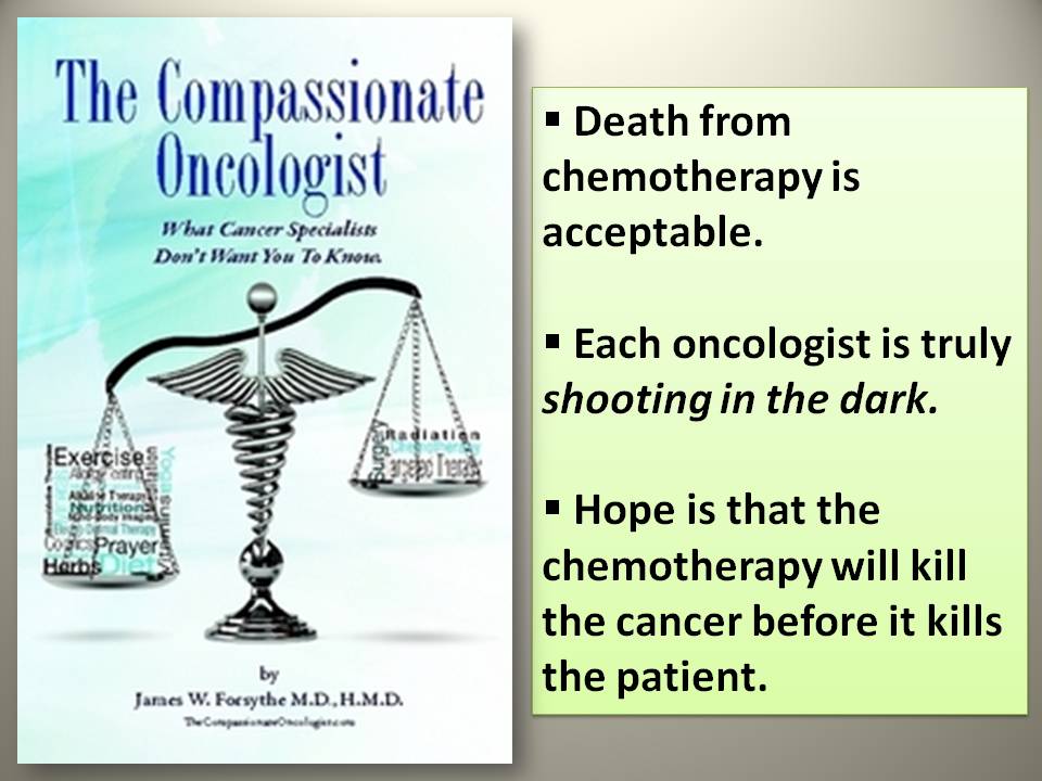
Today, many drugs similar to angiostatin (such as Avastin, Sutent and Nexavar) have been developed by the pharmaceutical industry. But “their effect on humans when used alone have turned out to be disappointing” (ANTICANCER page 54). This view is also shared by medical oncologist Dr Richard Frank, MD (in FIGHTING CANCER WITH KNOWLEDGE AND HOPE) in which he said that…“although targeted therapies (angiogenesis inhibitor drugs as mentioned above) were developed with the hope that they would be magic bullets that would neatly eradicate cancer through the selective targeting of one critical molecule, in general they have fallen short of their lofty goal” (page180). Anti-angiogenesis drugs have produced more troublesome side effects than foreseen. As a result, they are probably not the long-hoped-for miracle drugs (ANTICANCER page 54).

According to Dr David Servan-Schreiber, as an alternative to waiting for the miracle drug, there are natural approaches that have a powerful effect on angiogenesis without side effects and that can be combined perfectly with conventional treatments (page54). These are:

  • Specific dietary practices (many natural anti-angiogenesis foods have been discovered recently, including common edible mushrooms, green tea, spices, and herbs)..
  • Everything that contributes to reducing inflammation, the direct cause of the growth of new blood vessels.

Anti-angiogenesis foods listed by Dr Schreiber are green tea, olives and olive oil, turmeric and curry, ginger, cruciform vegetables, garlic, onion, leeks, shallots, chives, vegetables and fruits rich in carotenoids, tomatoes and tomato sauce, soy, mushrooms, herbs, and spices, seaweed, berries, plums, peaches & nectarines, citrus fruits, pomegranate juice, red wine, dark chocolate, vitamin D, Omega-3s, probiotics and foods rich in selenium. (For a complete exposure of these foods we urge you to read Chapter 8: The Anti-Cancer Foods. We also urge you to watch the DVD entitled “AntiCancer with Dr David Servan-Schreiber.” Some links are available on YouTube.com as follows:

a)   Dr David Servan-Schreiber’s Remarkable Story:

http://www.youtube.com/watch?v=xfddD6keYq0

b)   Natural Defences in Preventing and Treating Cancer:

http://www.youtube.com/watch?v=XaDt3AJQ98c 

Anti-angiogenis or anti-angiogenic foods? Your doctor/ oncologist will in all probability pour scorn on this concept with the usual comments–not proven, not scientifically tested, etc. But frankly, are all the conventional cancer treatments properly and scientifically and independently tested?

Who else has done research and written about anti-angiogenic dietary factors under the concept of angiogenesis?

In the forefront of such research is Dr William Li MD, the founder of The Angiogenesis Foundation, the world’s first non-profit organisation dedicated to conquering disease using the new approach based on angiogenesis, the growth of new capillary blood vessels in the body.

According to Dr Li, many foods contain naturally occurring inhibitors of angiogenesis. When these foods are consumed and absorbed into the blood stream, the inhibitors act to boost the body’s existing system that suppresses undesirable angiogenesis that can promote or accompany disease.

The following is a list of foods (according to Dr Li) that have innate properties which inhibit angiogenesis, thus working to cut off cancer tumours from blood supplies. These are green tea, berries, citrus fruits, apples, pineapple, cherries, red grapes, red wine, cruciferous vegetables, soybeans, ginseng, mushroom, liquorice, turmeric, nutmeg, lavender, artichokes, pumpkin, sea cucumber, tuna, parsley, garlic, tomato, olive oil, grape seed oil, dark chocolate. (Source: Angiogenesis Foundation Website: http://www.angio.org).

Also we recommend that you watch a video of Dr William Li enlightening you about “angiogenesis,” its impact on the human body, its connection to cancer and how you can deal with it.

To view the video, try the following links: –

a)    http://www.youtube.com/watch?v=C_5Z31mUmtc

  • or just type in Dr William Li on YouTube or on Google 

Dr Judah Folkman’s visionary ideas on cancer treatment served as a starting point and inspired two Canadian cancer researchers to theorise and confirm that “there is some weakness in the armor of tumor cells that might allow us to better our chances of destroying them” (Incidentally Chapter 4 in Dr Schreiber book “ANTICANCER” is entitled “Cancer’s Weakness”) These two researchers Dr Richard Beliveau, PhD and Dr Denis Gingras, PhD worked on the premise that “despite its great power, its versatility, and its enormous ability to adapt to hostile conditions of neighbouring cells, the cancer cells remains extremely dependent upon its energy needs. To grow, a tumour requires a constant supply of oxygen and nutrients. Their studies strongly suggest that certain types of cancers can be prevented by modifying our dietary habits to include foods with the power to fight tumours at the source and thus prevent their growth.

According to Dr Believeau and Dr Gingras, “nature supplies us with an abundance of foods rich in molecules with very powerful anticancer properties capable of engaging with the disease without causing any harmful side effects. In many respects, these foods possess therapeutic properties on par with those of synthetic drugs” (Ha, Big Pharma definitely won’t like this statement)

Some of the specific foods researched by Dr Beliveau and Dr Gingras are: cruciferous vegetables, garlic and onions, soy, turmeric, green tea, berries, omegs-3s, tomatoes, fresh fruits, and dark chocolates.

Dr Beliveau and Dr Gingras distilled their research findings into a simple book for the lay person- “FOODS TO FIGHT CANCER” –the goal of this book is to present a summary of the scientific studies currently available.

Another medical doctor who believes and has written on the subject of angiogenesis is Dr Joel Fuhrman, a board-certified family physician who specializes in preventing and reversing disease through nutritional and natural methods. In this book “SUPER IMMUNITY” Dr Fuhrman touched on angiogenesis in Chapter 3 under the heading, “The Anticancer Solution” The salient points in this section are: –

  • Many plant foods contain natural angiogenesis inhibitors- especially mushrooms
  • Dietary angiogenesis inhibitors are now being investigated as a preventive strategy to “starve” cancers while they are still small and harmless.
  • If our diet contains plenty of angiogenesis inhibitors, it can prevent small tumours from acquiring a blood supply and growing larger and becoming more aggressive or cancerous.
  • Some anti-angiogenic foods/nutrients listed by Dr Fuhrman are allium vegetables, berries, black rice, cinnamon, citrus fruits, cruciferous vegetables, flax seeds, ginger, Grapes, green tea, mushrooms, Omega-3 fats, peppers, pomegranate, quince, resveratrol, soybeans, spinach, tomatoes, and turmeric. (Scientific studies are quoted by Dr Fuhiman in the end NOTES)
  • On the other hand, “there are foods and nutrients that promote angiogenesis–and thus obesity and cancer. These include white-flour based breads and sweets that raise insulin levels, and the high-fat, high-cholesterol, standard, Western diet. These modern, unhealthy foods promote fat storage in addition to having a high-caloric density. They are a double negative, while green, mushrooms, onions, berries and the other foods listed above are a double positive”

In concluding the chapter, Dr Fuhrman laments that… “many people choose to reject new science even when the evidence is overwhelming. This book, SUPER IMMUNITY, may be attacked by people in powerful positions of authority whose livelihood is dependent on competing interests such as “recreational” foods, drugs and medical technology. Does this sound familiar to you?

In “FIGHTING CANCER WITH KNOWLEDGE AND HOPE,” oncologist Dr Richard Frank clearly stressed that:

  • Diet can promote or inhibit the formation of cancer in many ways
  • There are both good and bad foods to influencing the development of cancer
  • More direct links between particular components of food and cancer have been confirmed by some recent studies. A classic link is attached.

Link: http://www.ncbi.nlm.nih.gov/pubmed/17699009

Although “anti-angiogenesis drugs (like Avastin, Sutent, Nexavar) prevent tumours from growing the blood vessels they need to grow, none is perfect” (page 481). This is the view of Dr Keith Block, MD an Integrative Oncologist who explained that “just as tumours can switch to a second growth pathway if their primary pathway is blocked by a chemotherapy drug, so tumour can switch to a backup pathway for growing blood vessels when the first pathway is blocked by an anti-angiogenesis drug”(page 481).

Just as drug cocktails are a hot area of research in mainstream oncology, so combinations of anti-cancer compounds are some of the most exciting advances in integrative care…. there exists natural compounds that target the same growth pathways as leading-edge pharmaceuticals (page 505).

Some of natural compounds that have anti-angiogenic properties are berries (most types) which inhibit production of VEGF, a common growth pathway, and also prevent angiogenesis. The soy compound genistein also inhibits VEGF and angiogenesis which may be one reason soy is associated with lower cancer rates. Other natural compounds that can stimulate cells of the immune system to seek out and identify malignant cells are: aloe vera, acemannan, ginseng, curcumin, green tea polyphenols, resveratrol, mushrooms, grape seed extract, etc. (page 505/507)

All the above comments by Dr Block are contained in his bestselling book “LIFE OVER CANCER” which we recommend that you read the whole book or at least chapter 4 “The Anti-Cancer Diet” In this chapter, you will learn why you should not eat the following when you have cancer: –

  • Animal Protein
  • Bad Fats
  • Refined Carbohydrates
  • Dairy Products

Dr Block strongly believes that diet affects cancer both directly and indirectly. Nutrients directly impact the mechanisms by which cancer cells grow and spread. They indirectly help control the cancer by changing the surrounding biochemical conditions that either encourage or discourage the progression of malignant disease. The bottom line is that what you eat can spell the difference between conquering your disease or having it rage out of control (page 56).

For more information of the book by Dr Block, visit the following links:

a)    http://www.lifeovercancer.com/

b)   http://lifeovercancerblog.typepad.com/

Dr Margaret Cuomo, MD, and a board–certified radiologist wrote the book, “A WORLD WITHOUT CANCER” gave a few tips on “Fighting Cancer with Nutrition and Physical Activity.” Dr Cuomo suggests the following for a Cancer-Prevention Diet: –

a)    Eat more fruits and vegetables – such as berries, cruciferous vegetables, tomatoes, dark green, leafy vegetables (page 205).

b)   Buy organic – The International Agency for Research on Cancer classifies more than 400 chemicals, including those used in pesticides, as carcinogens (page 206).

c)    Eat more Fibre – fibre dilutes the carcinogens in the colon; reduce the time in which they remain there, enhanced anti-oxidant action, or produce bacteria that promote, or produce bacteria that promotes a healthy digestive tract (page 206).

d)   Avoid Red Meat – a growing body of evidence points to an association between beef, pork, lamb, and goat and cancers of the colon, prostate, pancreas and kidney (page 208/209). Carcinogens may also be present in smoked, salted, or cured meat and in meats cooked at high temperatures.

Besides the above, Dr Cuomo also advise cancer patients to eat more fish, drink green tea, increase consumption of resveratrol, flavor food with turmeric and lastly to limit processed foods (page 207-209).

For further reference, read Dr Cuomo’s article:

  • Cancer Prevention Tips from Dr Margaret Cuomo, MD

Link : http://blog.tjmartell.org/cancer-prevention-tips-from-dr-margaret-cuomo/

Another prominent medical doctor, Dr Russell Blaylock, a board-certified neurosurgeon, believes that “nutrients do block angiogenesis” (pages 182/183)….especially the flavonoids from edible plants such as genistein extracted from soybeans, catechins found in grape-seed extracts, apigenin and luteolin which occur in high concentrations in celery. In his book, “NATURAL STRATEGIES FOR CANCER PATIENTS,” Dr Blaylock advised that doing two things will significantly reduce tumour angiogenesis:

  • Correcting your dietary ratio of omega-6 and omega-3 fats,
  • Increasing your intake of vegetables.

Essentially, it means that a diet of omega-3 products inhibits angiogenesis and a diet high in the omega-6 fats powerfully promotes cancer growth and spread. Nicotine also increases angiogenesis.

A prominent cancer researcher and scientific advisor to the University of Texas Centre for Alternative Medicine, D John Boik, PhD is the author of 2 very scientific texts……CANCER AND NATURAL MEDICINE and NATURAL COMPOUNDS IN CANCER THERAPY. In the 2 books, the subject of angiogenesis is extensively covered.

Some of the natural inhibitors of angiogenesis are curcumin, EPA and DHA, garlic, melatonin, resveratrol, plant flavanoids (genistein, apigenin, luteolin, quercetin, green tea catechins such as EGCG). Read Chapter 8-Natural Inhibitors of Angiogenesis. In this chapter, Dr Boik also pointed out that…”eicosanoids derived from omega-6 fatty acids facilitate cancer progression and eicosanoids derived from omega-3 fatty acids inhibit it.”

Finally, we would like to share with you an E-Book or Nook Book that we found and it is written by Dr Hratch Karamanoukian, MD and a prominent cardiovascular and thoracic surgeon who has specialized in minimally invasive cardiac surgery, thoracic surgery, robotic surgery and vein disorders. In 40 FOODS THAT FIGHT CANCER,” he shares his wisdom as follows:

  • Some foods can help you to fend off cancer, while others could actually be increasing your risk of cancer. Knowing the right foods to add to your diet is very important.
  • Choosing the best foods will be able to help you strengthen and build your immune system, which means fighting off diseases is going to be easier. The right foods are going to make your body stronger and increase your overall health

The following are the 40 Foods that Dr Karamanoukian recommends in his book:

  • Eat more vegetables……broccoli, cabbage, cauliflower, kale, mushrooms, seaweed, sweet potatoes, turnip greens, onions, summer and winter squash, spinach, olives and Brussels sprouts.
  • Add more fruits to your diet…..tomatoes, avocadoes, grapefruit, figs, oranges, papaya, raspberries, blueberries, strawberries, pears, grapes and lemons.
  • Spices, beans and other foods to help fight cancer…..garlic, sunflower seeds, oregano, turmeric, red wine, peanuts, ginger, tea, brown rice, black beans, ground flaxseed, quinoa, peppermint and fish.

BESIDES THE ABOVE BOOKS REVIEWED, YOU MAY WISH TO READ FURTHER. WE RECOMMEND THE FOLLOWING LINKS:

a)    THE ANGIOGENESIS FOUNDATION    http://www.angio.org/

On the main page, click on UNDERSTANDING ANGIOGENESIS and then click on DIET, LIFESTYLE AND ANGIOGENESIS. 

b)   EAT TO DEFEAT CANCER  http://www.eattodefeat.org/

On the main page, click on Food to view the list of foods profiled as cancer-fighting foods and then click on Evidence for a list of articles to read.

c)    www.doctoroz.com   http://www.doctoroz.com/videos/stop-cancer-growing

d)   AG SCIENTIFIC BLOG

Part 1: http://info.agscientific.com/blog/bid/132253/Eat-For-Your-Life-Top-Anti-Angiogenesis-Foods-Part-1

Part 2: http://info.agscientific.com/blog/bid/133114/Eat-For-Your-Life-Top-Anti-Angiogenesis-Foods-Part-2 

e)    www.mercola.com    http://articles.mercola.com/sites/articles/archive/2010/06/08/dramatically-effective-new-natural-way-to-starve-cancer-and-obesity.aspx

There are a lot more of other such articles…..just google for either anti-angiogenesis or anti-angiogenic foods.

After you have read this far, you would definitely be able to differentiate between foods that inhibit angiogenesis and foods that promote angiogenesis. Remember, your life is in your hands….not in your doctor’s and the choice is yours to decide. 

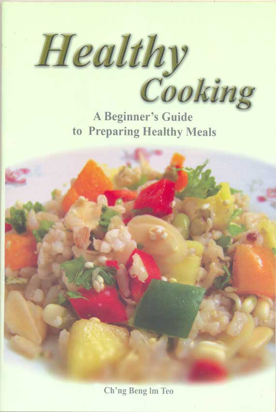
NB: If you are still unsure as to what to cook or how to cook, get hold of a copy of HEALTHY COOKING …A Beginner’s Guide to Preparing Healthy Meals by Ch’ng Beng Im Teo. (ISBN NO: 978-983-2590-25-5).

1 Cover


NB: THESE NOTES, COMPILED BY YEONG SEK YEE AND KHADIJAH SHAARI, ARE MEANT STRICTLY FOR YOUR INFORMATION AND NOT INTENDED TO DISSUADE YOU FROM SEEKING CONVENTIONAL CANCER TREATMENTS. THIS HAS TO BE SOLELY YOUR RESPONSIBILITY/DISCRETION.


Kidney Cancer: An expensive and dangerous drug that does not cure

KC (E87) is a 50-year-old male. In late January 2013, he had breathing difficulties and had to be hospitalized.  There was fluid in his lung. Pleural tapping was done. KC’s health was restored after 10 days in the hospital where 4 litres of pleural fluid was tapped out.

A CT scan done on 21 February 2013 indicated the following:

  1. Multiple pulmonary nodules in both lower lobes. Right pleural effusion and enhancing right pleural nodules.
  2. Bilaateral heterogeneously enhancing renal masses in upper pole measuring 7.3 x 6.6 cm (right) and 7.0 x 8.1 cm (left).
  3. Right lobe liver with ill defined lesion in segment IV of liver suggestive of local infiltration.
  4. Multiple enhancing peritoneal nodules in right subhepatic space.
  5. Enlarge retrocrural lymphadenopathy and multiple subcentimeter paraaortic and aortocaval nodes. Mild to moderate ascites.

Impression:  Bilateral renal masses likely renal cell carcinoma with local infiltration and lung pleural and peritoneal metastases.

After the CT scan KC was asked to go home and nothing was done. There was no medication either. No satisfied, KC went to consult an oncologist at a private hospital. Another CT scan was done on 26 March 2013. The results indicated:

  1. Multiple mildly enlarged mediastinal nodes.
  2. There is a small-to-moderate right pneumothorax (a collapsed lung – a collection of air in the space around the lungs. This buildup of air puts pressure on the lung, so it cannot expand as much as it normally does when taking a breath).
  3. Pleural mass at the right anterior costophenic sulcus measuring 8 x 5.6 x 3.8 cm.
  4. Another large right pleural based mass noted anteriorly underlying the right third rib and it measures 6 x 2.7 x 2 cm.
  5. There is also a fairly large pleural based mass medially abutting the left atrium. It measures 5 x 4 x 3 cm.
  6. Bilateral multiple slightly lobulated pulmonary nodules with two nodules at the left lung apex demonstrating cavitation.
  7. There are bilateral large heterogenous masses occupying the upper half of both kidneys. The right renal mass measures 9 x7.4 x 6.7 cm. The left renal mass measures 8.7 x 8 x 6.6 cm.
  8. There is a peritoneal heterogenous lesion invading segment 6 of the liver, measuring 5.6 x 5 x 5 cm.
  9. There are multiple mildly enlarged retroperitoneal nodes measuring up to 17 mm.
  10. There is biconcave appearance of T3 vertebral body due to compression fracture.
  11. There is deformity at the left iliac crest, due to previous bone grafting.

KC was told by the oncologist that since both his kidneys are infected with cancer, surgery is not indicated. The cancer has also spread to his lungs and liver. The oncologist prescribed Sutent. The total cost of his first visit to the oncologist was RM 7,000. Sutent itself cost RM 5,000 for 9 days consumption. The remaining RM 2,000 was for the CT and RM 300 for consulting the oncologist.

KC came to seek our help on 5 April 2013. The following are excerpts of our conversation.

Chris: Did they remove your kidneys?

KC: No, both my kidneys were infected. They could not remove them.

C: Then you saw the oncologist, what did he do?

KC: He prescribed me this drug (showing Sutent box).

C: How much did this cost you?

KC: RM 5,000 for 9 days of consumption.

C: Did you ask if the drug is going to cure you?

KC: Only to control the spread of the cancer.

C: How long do you need to take this drug?

KC: He cannot tell me. I told the doctor. This drug is expensive, I cannot afford it. He said: How much is your life worth? And when I talked too much he got angry at me. I told him I cannot afford to take this medication. I only earn RM 1,000 plus per month – how to afford it.

C: Did he tell you the adverse side effects of his drug?

KC: ?? No, I told him I cannot afford to take the medication. He asked me: How much is your life worth? And why do you come and see me then?

Comments:  This is indeed a sad and tragic story.  Just reflect on this: How long do you expect a patient who earns RM 1,000 plus per month afford to take drug that cost him RM 15,000 per month?  What is the rationale of asking this patient to take the drug? To cure him or is this for any other purpose?  Patient was to take Sutent so as to stop the cancer from spreading. This unfortunately is the line very often sold to patients to make things attractive. But is that an honest answer based on scientific fact? Read below and you will be shocked!

This is something you need to know that oncologists or those with vested interests, will not tell you!

Sutent-spread-cancer 2

Sutent box

Read more: http://www.dailymail.co.uk/health/article-2088032/Cancer-drugs-aim-shrink-tumours-cutting-blood-supply-actually-help-SPREAD.html#ixzz2PwQx0Tun

  • Cancer drugs that shrink tumours by cutting off their blood supply may end up helping them to spread, a study suggests.
  • Tests on mice showed that both Glivec and Sutent depleted pericytes by 70 per cent while metastasis rates tripled.
  • Glivec, the brand name of the drug imatinib, and Sutent (sunitinib) have both been shown in trials to increase patient survival by a significant degree.
  • Drugs such as Glivec and Sutent reduce the size of tumours but could also make them more aggressive and mobile.
  • As a result tumours find it easier to ‘metastasise’, or spread around the body.

If you just looked at tumour growth, the results were good,‘said lead researcher Professor Raghu Kalluri, from Harvard Medical School in Boston. But when you looked at the whole picture, inhibiting tumour vessels was not controlling cancer progression. The cancer was, in fact, spreading. 

Seventy to 80 percent of cells in breast tumour are non-cancer cells. Are they all bad? Some of them are there to protect us and they’re not all bad.

Indeed  Sutent has been approved by the US FDA for treatment of advanced renal cancer, but the reality is it does not cure advanced or metastatic renal cancer or any cancer at all for that matter. Know that!

Below is the Information obtained from the drug company’s website:  http://www.sutent.com/rcc.aspx

The website said, Sutent has been proven effective in the treatment of advanced RCC (renal cell carcinoma).

A clinical study has proven that Sutent is more effective than interferon alfa (IFNα), another approved treatment option for advanced RCC.

SUTENT results:

  1. SUTENT More Than Doubled Median Progression-Free Survival (PFS) vs IFNα – 11 months vs 5 months with IFNα. 
  2. SUTENT Achieved More Than 2 Years’ Median Overall Survival (OS):  26.4 months vs 21.8 months with IFNα.
  3. FIVE TIMES more people saw their tumors shrink. 103 patients taking Sutent compared to 20 taking IFNα. 

Of course, when you read this information it is very impressive (but don’t fall into the trap of sweetened or massage research data!). Why do I say the message is a sweet camouflage?  Let’s look at the data critically.

  1. If you take Sutent, you have TWO TIMES longer progression-free survival.   Progression-free survival means the length of time from the start of treatment that patients remain alive and their disease does not worsen. The question you want to ask is:, what if you don’t take Sutent – can you also have progression free survival? Of course, but it is shorter a bit. According to the Drug Company, with Sutent the progress-free survival was 11 months compared to 5 months without Sutent. So Sutent gave an advantage of 6 months.Progression-free does not mean cure at all. It is just the disease does not worsen and you are still alive.
  2. Those who take Sutent remained alive. The median overall survival was 26.4 months and then they die. But what if you don’t take Sutent? Patients were still alive but died at 21.8 months. That means by taking Sutent you live longer by 4.6 months.

Take note:  you live longer by only 4.6 months. Is that what you want? Let us look at the economics. To live 4.6 months longer you need to take Sutent for at least 27 months, according to this study. This would cost you about RM 15,000 x 27 = RM 405,000. I am sorry, is my calculator playing tricks on me? Is that not a lot of money to you and me? And spending that kind of money to chase after 4.6 months of extended life? Let us hope that the extra 4.6 months of life is blissful, worth every minute of it. Or is it? There is no free lunch – you may have to suffer severe side effects as listed below.Nowhere in medical literature do I ever come across data showing that Sutent cures your cancer.

3. Five times as many people on Sutent saw their tumors shrink – is this a great achievement? Shrinkage of tumour has NO meaning. It is just a lure or camouflage! And what do you make out of the research led by Professor Raghu Kalluri of Harvard Medical School in Boston which said that Sutent may shrink the tumour but ultimately Sutent  may end up helping cancer spread more aggressively and widely.

The following are warnings given by the Drug Company.

SUTENT may cause serious side effects, including:

  • Serious liver problems, including death
  • Heart problems—include heart failure and heart muscle problems (cardiomyopathy) that can lead to death.
  • Abnormal heart rhythm changes— you feel dizzy, faint, or have abnormal heartbeats
  • High blood pressure.
  • Bleeding sometimes leading to death— these symptoms may include:
    • Painful, swollen stomach (abdomen)
    • Bloody urine
    • Vomiting blood
    • Headache or change in your mental status
    • Black, sticky stools
    • Jaw-bone problems (osteonecrosis)—severe jaw bone problems may happen.
    • Tumor lysis syndrome (TLS)— caused by the fast breakdown of cancer cells and may lead to death. TLS may cause nausea, shortness of breath, irregular heartbeat, clouding of urine and tiredness associated with abnormal laboratory test results (high potassium, uric acid and phosphorous levels and low calcium levels in the blood) that can lead to changes in kidney function and acute kidney failure.
    • Hormone problems, including thyroid and adrenal gland problems— as in the  following signs and symptoms:
      • Tiredness that worsens and does not go away
      • Heat intolerance
      • Loss of appetite
      • Feeling nervous or agitated, tremors
      • Nausea or vomiting
      • Sweating
      • Diarrhea
      • Irregular menstrual periods or no menstrual periods
      • Fast heart rate
      • Headache
      • Weight gain or weight loss
      • Hair loss
      • Feeling depressed

Common side effects of SUTENT include:

  • It may make your skin look yellow. Your skin and hair may get lighter in color
  • Tiredness
  • Weakness
  • Fever
  • Gastrointestinal symptoms, including diarrhea, nausea, vomiting, mouth sores, upset stomach, abdominal pain, and constipation.
  • Rash or other skin changes, including drier, thicker, or cracking skin
  • Blisters or a rash on the palms of hands and soles of feet
  • Taste changes
  • Loss of appetite
  • Pain or swelling in arms or legs
  • Cough
  • Shortness of breath
  • Bleeding, such as nosebleeds or bleeding from cuts

How much is your life worth?

Let me ask you to ponder what Rabbi Harold Kushner said:

11-Kushner-Moral-compass

Perhaps we all should read the article below presented by Drs Tito Fojo and Christine Grady. The first author is from the Medical Oncology Branch of the National Cancer Institute, Bethesda, USA, while Dr. Grady is from the Clinical Center, National Institutes of Health, Bethesda, USA.

Sutent-How-much-is-life-wor

Read more: http://jnci.oxfordjournals.org/content/101/15/1044.full.pdf+html

Read also https://cancercaremalaysia.com/2011/05/27/dissecting-chemotherapy-part-4-how-much-is-life-worth-erbitux-for-lung-cancer/

In their paper, Dr. Fojo & Grady wrote:

  • The all too common practice of administrating a new, marginally beneficial drug to a patient with advanced cancer should be strongly discouraged.
  • In cases where there are no further treatment options, emphasis should be first on quality of life and then cost.
  • For therapies with marginal benefits, toxic effects should receive greater scrutiny.
  • We must deal with escalating price of cancer therapy now.
  • The current condition cannot continue … the time to start is now.
  • As oncologists, we cannot go without answering these questions. The moral character of our specialty depends on the answers.

You may wish to read the following related stories:

  1. Kidney-Lung-Brain Cancer: Sutent = Heart Damage https://cancercaremalaysia.com/2011/09/19/kidney-lung-brain-cancer-sutent-heart-damage/
  2. Sutent for Advanced Kidney Cancer  https://cancercaremalaysia.com/2011/09/18/sutent-for-advanced-kidney-cancer/
  3.  Kidney Cancer Part 2: Two Oncologists Two Different Opinions – Is Sutent indicated in this case? https://cancercaremalaysia.com/2011/09/19/kidney-cancer-part-2-two-oncologists-two-different-opinions-%E2%80%93-is-sutent-indicated-in-this-case/
  4. Die of Cancer But Don’t Die A Bankrupt! https://cancercaremalaysia.com/category/kidney-cancer/

 More:

Perilous Approach: Avastin and Sutent Promote Growth of Breast Cancer Stem Cells

Avastin

(Note: Many patients in Malaysia have been treated with this very expensive drug)

http://badscienceblindtruth.wordpress.com/2013/04/19/perilous-approach-avastin-and-sutent-promote-growth-of-breast-cancer-stem-cells/

Medical Bankruptcy in the US

After reading Steven Brill article, Bitter Pill: Why medical bills are killing us (Time magazine, 20 February 2013) I started to surf the Internet and posed some questions. Let me share with you what I found.

Frederick Allen of Forbes wrote an article: The Reason American Health Care Is Out Of Control http://www.forbes.com/sites/frederickallen/2013/03/05/the-reason-american-health-care-is-out-of-control/

This is what he wrote:

Steven Brill’s Time magazine cover story last week on health care got a lot of attention for its tracking of astronomical costs in American health care back to their sources. But why are those costs so high anyway? Is it because an unfettered free market is not allowed to work when it comes to health and medicine? Quite to the contrary.  It’s partly because an unfettered free market cannot work when it comes to health and medicine … a free market can’t control those costs. It can only distort and encourage them. It makes them worse. And it isn’t even really a free market.

As Steve Brill wrote: Everyone along the supply chain—from hospital administrators (who enjoy multimillion-dollar salaries) to the salesmen, executives and shareholders of drug and equipment makers—was reaping a bonanza.

And the only free-market choice I could make at this point to bring way down my own astronomical medical costs would be the choice that was so famously given to Jack Benny: Your money or your life.

Two readers responded to Frederick Allen’s article:

  • Doctors in the US have a very powerful cartel restricting the supply of new doctors – medical school slots and residency positions are not increasing due to the demographic-driven demand. Mid-level providers (Nurse Practitioners and Physician Assistants) help meet the demand; but the barriers to becoming a doctor can’t help control prices.
  • It is very true that people shall pay anything for preserving life! This very compulsion of a patient, is exploited by everyone in the medical sector where there exists nexus between doctors and insurance companies on one hand and on the other hand another nexus exists between pharmaceutical companies and medical clinics/doctors. In their greed to milk a needy patient, every means is employed to make him pay for extremely exorbitant prices for medicines even though cheaper substitutes could be available. There must be some accountability in such matters. Medical profession has turned out to be an extremely lucrative profession in the present times. If patients are left a choice to shop for buying the prescribed medicines from adjoining Canada or Mexico, they would get exactly same named prescribed medicine, manufactured by the same company, at a much cheaper rate over there. The US. Federal government rather should give a choice to those patients who can procure those prescribed medicines at cheaper rates from Canada/Mexico etc.

I typed this question in Google search: Is the US healthcare cost-effective?

The US healthcare system is one of the least cost-effective in reducing mortality rates; while the United Kingdom is among the most cost-effective, according to a recent study published in the July issue of Journal of the Royal Society of Medicine Short Reports. The study compared the United States, United Kingdom, and 17 Western countries’ efficiency and effectiveness in reducing mortality over a 25-year period. The greatest cost-effectiveness  was found for Ireland, United Kingdom, and New Zealand  and the  least cost effective, were found for Portugal, Switzerland, and the United States.

Read more: http://formularyjournal.modernmedicine.com/news/us-healthcare-system-among-least-cost-effective-reducing-mortality

Umair Haque, Director of Havas Media Labs and author of Betterness: Economics for Humans  wrote:

  • Unless you’ve been living under a rock, you’ve heard by now that, where the majority of developed countries spend between 8–10% of GDP on health, America spends ~16%. Per capita healthcare costs in the States have significantly outstripped costs in other countries.
  • Why has healthcare expenditure exploded? Each component of healthcare spending has grown — but the fastest growth has come from prescription drug spending. Where people in other developed countries spend between $400 and $500, Americans spend almost $900 per capita on pharmaceuticals.
  • Americans receive less care than their counterparts in other developed nations. The US has the lowest number of hospital beds per 1,000 people amongst developed countries. And it has the smallest number of doctor consultations per capita — just 3.8, compared to Canada’s 5.8, or Germany’s 7.4.
  •  Americans pay more for healthcare because they trade more expensive products for less service, realizing poorer outcomes. Why? Because that is what maximizes near-term profits along the value chain.

1-Profit-of-pharma

 Read more: http://blogs.hbr.org/haque/2009/08/how_to_think_constructively_ab.html

I typed this question in Google search: Is the US healthcare compassionate?

Rick Nauert, in an article: Compassion Missing in American Health Care wrote: Compassionate care is defined by the following four essential characteristics:

1. Empathy, emotional support, and a desire to relieve a patient’s distress and suffering.
2. Effective communication at all stages of a patient’s illness and treatment.
3. Respecting patients’ and families’ desires to participate in making health care decisions.
4. Knowing and relating to the patient as a whole person, not just a disease.

Compassion is as important in helping patients manage chronic and acute conditions as it is at the end of life. To improve quality and reduce costs, compassion should be present in all aspects of our healthcare system.

The survey found that only 53 percent of patients and 58 percent of doctors rate the U.S. healthcare system as a compassionate one.  Problems with the U.S. health care system include escalating costs, medical errors, inconsistent results and, according to a new national survey, a lack of compassion.

Read more: http://psychcentral.com/news/2011/09/09/compassion-missing-in-american-health-care/29295.html

Gordon Marino, professor of philosophy at St. Olaf College  wroteThe real US healthcare issue: compassion deficiency. Americans suffer from a compassion deficiency.

Read more: http://www.csmonitor.com/Commentary/Opinion/2009/0813/p09s01-coop.html 

I typed this question in Google search: Is the US healthcare money driven?

There is a movie –  Money-Driven Medicine –  produced by Academy Award winner Alex Gibney (Taxi to the Dark Side, etc.) and inspired by Maggie Mahar’s acclaimed book, Money Driven Medicine: The Real Reason Health Care Costs So Much.  The message of this movie:

  • The U.S. spends twice as much per person on healthcare as the average developed nation,  yet our outcomes, especially for chronic diseases, are very often worse.
  • What makes us the exception? The U.S. is the only industrialized nation that has chosen to turn medicine into a largely unregulated, for-profit business.
  • Dr. Donald Berwick, Administrator of the Centers for Medicare and Medicaid Services, explains: We get more care, but not better care.
  • Medical ethicist Larry Churchill doesn’t mince words: The current medical care system is not designed to meet the health needs of the population. It is designed to protect the interests of insurance companies, pharmaceutical firms, and to a certain extent organized medicine. It is designed to turn a profit. It is designed to meet the needs of the people in power.
  • As the eye-opening ads in Money-Driven Medicine reveal, the more new drugs, surgical procedures, diagnostic devices and hospital beds the health industry can produce, the more they can sell – whether we need them or not. It’s called “supply-driven demand” and it’s possible because a sick consumer can’t say no.

Read more: http://moneydrivenmedicine.org/about-mdm

In another article: 50 Signs That The U.S. Health Care System Is A Gigantic Money Making Scam That Is About To Collapse, the author wrote:

  • The U.S. healthcare system is a giant money making scam that is designed to drain as much money as possible out of all of us before we die.
  • In the United States today, the healthcare industry is completely dominated by government bureaucrats, health insurance companies and pharmaceutical corporations.  The pharmaceutical corporations spend billions of dollars to convince all of us to become dependent on their legal drugs, the health insurance companies make billions of dollars by providing as little health care as possible, and they both spend millions of dollars to make sure that our politicians in Washington D.C. keep the gravy train rolling.
  • Healthcare costs continue to go up rapidly, the level of care that we are receiving continues to go down, and every move that our politicians make just seems to make all of our healthcare problems even worse.
  • In America today, a single trip to the emergency room can easily cost you $100,000, and if you happen to get cancer you could end up with medical bills in excess of a million dollars.
  • Even if you do have health insurance, there are usually limits on your coverage, and the truth is that just a single major illness is often enough to push most American families into bankruptcy.  At the same time, hospital administrators, pharmaceutical corporations and health insurance company executives are absolutely swimming in huge mountains of cash.

Read more: http://theeconomiccollapseblog.com/archives/50-signs-that-the-u-s-health-care-system-is-a-gigantic-money-making-scam-that-is-about-to-collapse

 I typed this in Google search:  Are Americans going bankrupt due to medical bill?

David U. Himmelstein, MD; Deborah Thorne, PhD; Elizabeth Warren, JD; and Steffie Woolhandler, MD, MPH are from the Department of Medicine, Cambridge Hospital/Harvard Medical School, Department of Sociology, Ohio University and Harvard Law School. They published their study: Medical bankruptcy in the United States, 2007 in the American Journal of Medicine, http://www.pnhp.org/new_bankruptcy_study/Bankruptcy-2009.pdf


What did they say? In 1981, only 8% of families filed for bankruptcy due to serious medical problem. In 2001, the author’s study showed that at least 46.2% of all bankruptcies were due to medical problems. In 2007, 62.1% of all bankruptcies were because of medical problems.

The shocking discovery: Most medical debtors were well educated, own homes and had middle class occupations. Three quarters had health insurance.

What has gone wrong? High medical bills directly contributed to their bankruptcy. Many families with continuous coverage found themselves under-insured, responsible for thousands of dollars in out-of-pocket costs. Others had private coverage but lost it when they became too sick to work. Nationally, a quarter of firms cancel coverage immediately when an employee suffers a disabling illness; another quarter do so within a year.

The authors concluded: The US healthcare financing system is broken.

CNN carried this report: Medical bills prompt more than 60 percent of U.S. bankruptcies. This year, an estimated 1.5 million Americans will declare bankruptcy. Many people may chalk up that misfortune to overspending or a lavish lifestyle, but a new study suggests that more than 60 percent of people who go bankrupt are actually capsized by medical bills. Bankruptcies due to medical bills increased by nearly 50 percent in a six-year period,

Read more: http://articles.cnn.com/2009-06-05/health/bankruptcy.medical.bills_1_medical-bills-bankruptcies-health-insurance?_s=PM:HEALTH

Reuters had this report: Medical bills underlie 60 percent of U.S. bankrupts: study. According to this report, medical bills are behind more than 60 percent of U.S. personal bankruptcies … healthcare reform is on the wrong track. More than 75 percent of these bankrupt families had health insurance but still were overwhelmed by their medical debts.

Dr. David Himmelstein of Harvard University said: Unless you’re Warren Buffett, your family is just one serious illness away from bankruptcy…For middle-class Americans, health insurance offers little protection.

http://www.reuters.com/article/2009/06/04/us-healthcare-bankruptcy-idUSTRE5530Y020090604

In the New York Times, Medical Bills Cause Most Bankruptcies, Tara Parket-Pope wrote:  Nearly two out of three bankruptcies stem from medical bills, and even people with health insurance face financial disaster if they experience a serious illness. The U.S. health care financing system is broken, and not only for the poor and uninsured,” the study authors wrote. “Middle-class families frequently collapse under the strain of a health care system that treats physical wounds, but often inflicts fiscal ones.

http://well.blogs.nytimes.com/2009/06/04/medical-bills-cause-most-bankruptcies/

Brennan Keller wrote in his blog: A new study done by Harvard University suggests that more than 62% of all personal bankruptcies are caused by the cost of over-whelming medical expenses. Of the most financially devastating diseases, cancer reigns supreme.

Read more: http://www.giveforward.com/blog/medical-expenses-top-cause-of-bankruptcy-in-the-united-states

Professor of Political Science, University of Missouri-St. Louis, Dr. Kenneth Thomas wrote: Medical Costs Help Drive United States to Highest Bankruptcy Rate in OECD.  A study published in the American Journal of Medicine shows that there was a sharp increase in the proportion of bankruptcies with significant medical causes (defined as debts over $5,000, loss of income due to health problems, or mortgaging of the debtor’s home to help meet medical expenses) between 2001 and 2007. According to their study, 46.2% of bankruptcies in 2001 were medically-related, while by 2007 the level had grown to 62.1%.

1-Bankcrupcy

http://middleclasspoliticaleconomist.blogspot.com/2012/02/medical-costs-help-drive-united-states.html

As expected, in any democratic country, there are always people who would dispute every scientific finding. Sally Pipes in her article: Medical bankruptcy: Fact or fiction?  http://thehill.com/blogs/congress-blog/economy-a-budget/263547-the-myth-of-medical-bankruptcy#ixzz2NHrAoqfD  wrote:

  • This year, a whopping 1.25 million Americans are expected to file for bankruptcy. Ask the president and his allies whom to blame, and they’ll point to healthcare. President Obama has claimed that the cost of healthcare causes a bankruptcy every 30 seconds.
  • But the alleged link between health costs and bankruptcy is about as real as the tooth fairy. The overwhelming body of research shows that medical costs play little or no role in the vast majority of U.S. personal bankruptcies.
  • Proponents of the health-cost-bankruptcy theory tend to cite a Harvard study that blames high medical bills for some 62 percent of American bankruptcies.
  • A study published in the journal Health Affairs reviewed Justice Department data and discovered that among Americans who cited medical debt as a contributing factor in their bankruptcy filing, only 12 to 13 percent of their total debts were medical.
  • The study also found that medical spending was a factor in no more than 17 percent of U.S. bankruptcies.
  • Too many Americans go bankrupt each year. But contrary to the claims … the cost of healthcare is not to blame. 

You can agree or disagree with what Sally Pipes wrote, but let me ask you to read again the article: Are medical bills killing patients?  $83,900 (approx: RM 251,700) the initial cost of a lymphoma treatment, or $902,452  (approx: RM 2.2 million) for treatment of lung cancer for 11 months before the patient died.

With that kind of medical bills, who would not go bankrupt?

As I surfed the internet further, I came across many shocking facts – I asked myself:  How not to go bankrupt with things like this happened?

The tab for medical care can add up quickly. Take a breast cancer diagnosis, for example.

  • A  breast cancer diagnosis bill can easily top $25,000.
  • A bilateral mammogram costs about $270.
  • A biopsy to test a suspicious area costs about $1,070.
  • A total mastectomy would cost about $11,500.
  • If the patient needs chemotherapy, a four-day hospitalization for treatment will run about $13,400.
  • Add another $260 per radiation treatment.

http://www.columbian.com/news/2012/nov/04/medical-bills-lead-many-families-to-file-for-bankr/

here are two interesting comments  in response to Towering Medical Bills Leave Many Americans Bankrupt:

  • This is written in loving memory of a couple I knew for decades. She suffered a lifetime with bi-polar. To pay for the expensive drugs not covered, he re-mortgaged. After retirement he could no longer keep up. The night before their foreclosure, he shot her and their border collie (dog) and out of grief torched their house. He is now serving life. The collie was killed “because he would have died of a broken heart”. Whenever I think of how inhuman our health care non-system is, my heart breaks again.
  • Our 25 year old son could not afford health insurance and it wasn’t provided at the restaurant where he worked. He had a serious accident which resulted in extensive third degree burns on his right hand, arm and leg. He was in the burn unit in Buffalo, NY for two weeks, underwent skin grafts and physical therapy to regain the use of his right hand and fingers. He ran up over 50K of medical bills but did not declare bankruptcy-because of the stigma. Now he is crippled by medical bills, cannot afford a car, his own apartment, or health insurance on his income. I don’t see how he will be able to recover from this and ever afford to have a family. 

Read more: http://www.npr.org/templates/story/story.php?storyId=105193107

  • When a Medicine (Revlimid) “Works” It’s Unaffordable: $132,000/year, $534/pill (This is RM 1,650 per pill) :  After making more than 70 phone calls to 16 organizations over the past few weeks, I’m still not totally sure what I will owe for my Revlimid, a derivative of thalidomide that is keeping my multiple myeloma in check. The drug is extremely expensive — about $11,000 retail for a four-week supply, $132,000 a year, $524 a pill. 
  • While drug companies spend a great deal to develop medications, their costs are inflated and overstated.  Efficiency is simply not on their agenda.  Nor is patient well being, access or for that matter outcomes. It’s the money stupid!  Charging more than twice the median U.S. income for a single drug that patients in life-threatening situation, any single drug, is simple blackmail.  It reflects an industry and economy gone mad. 
  • I had a friend who was on Revlimid for a pre leukemia condition. He was shocked when I told him what the cost was. The drug also has some very nasty side effects. Despite the treatment, he died a few months after starting it. 

Read more: http://medicynic.com/2012/12/09/and-when-a-medicine-revlimid-works-its-unaffordable-132000year-534pill/

  • Cabozantinib: A Miracle Cancer Drug without Survival Benefit:  The FDA recently approved cabozantinib for use in medullary thyroid cancer that has metastasized.  What’s noteworthy about this drug is that it will likely be very expensive and that it does not, repeat does not improve the patient’s survival – No statistically significant difference in overall survival.
  • Cometriq contains a Boxed Warning, telling doctors and patients about the risks of severe and fatal bleedings and perforations and fistula in the colon.
  • Other new miracle agents often have limited efficacy with say two months median survival improvement.  But this is the first such drug being actively promoted, that I can recall, that has no survival benefit. Maybe this is yet another reason we spend more on healthcare than any other country in the world.  

Read more: http://medicynic.com/2012/12/04/cabozantinib-another-miracle-cancer-drug-without-survival-benefit/

Read also: 

  1. The high cost of staying alive in a private hospital  https://cancercaremalaysia.com/2012/11/13/part-1-the-high-cost-of-staying-alive-in-a-private-hospital/
  2. Die of cancer but don’t die a bankrupt  https://cancercaremalaysia.com/2012/12/14/die-of-cancer-but-dont-die-a-bankrupt/
  3. How much life is worth  https://cancercaremalaysia.com/2011/05/27/dissecting-chemotherapy-part-4-how-much-is-life-worth-erbitux-for-lung-cancer/
  4. Money driven medicine  https://cancercaremalaysia.com/2011/09/02/book-review-money-driven-medicine-%E2%80%93-chemotherapy-for-non-responsive-cancers-%E2%80%93-denying-reality/
  5. Avastin does not cure cancer https://cancercaremalaysia.com/2011/05/27/dissecting-chemotherapy-part-6-avastin-does-not-cure-cancer/