Chemo and Tarceva Did Not Cure His Lung Cancer: Another meaningless decline of tumour markers

Jack (not real name) was 43 years old when he was diagnosed with lung cancer.  His problem started in October 2013, when there was a swelling in the right collar bone. Apart from this, there was no other symptoms — no cough, etc.  Jack said in 2013, he had fevers on three occasions that make him feel tired.

A blood test on 24 October 2013 showed his CEA was at 133.3 and CA 19.9 at 9,524.0

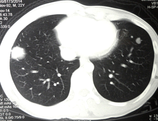
A CT scan on 30 October 2013 showed:

  • An irregular lesion in the right lung apex, 35 x 42 x 28 mm.
  • Speckles of calcification in both lungs upper lobe suggesting prior tuberculous infection.
  • Bilateral supraclavicular lymph node 10 to 20 mm.
  • Multiple enlarged mediastinal and right hilar lymph nodes.
  • Numerous small nodules in both lungs – likely metastases.
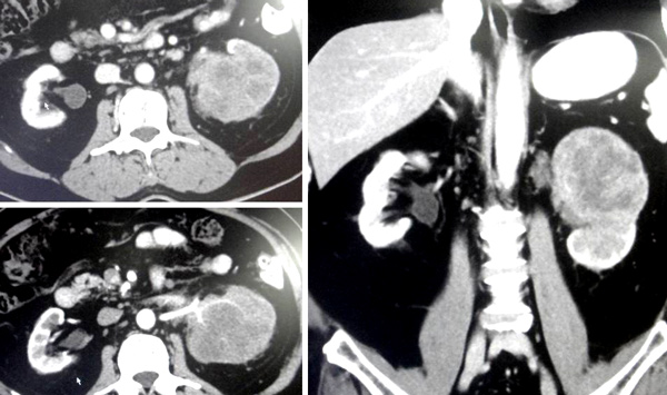
  • Liver shows a least 7 hypodense lesions, largest 30 x 32 mm in the left lobe – likely metastases.
  • Multiple enlarged lymph nodes in the celiac and paracaval regions.
  • Sclerotic lesions at vertebral bodies of L3, L4 and L5 – likely metastases.

A tru-cut biopsy of the right lung mass was performed and confirmed a moderately differentiated adenocarcinoma.

Jack underwent 6 cycles of chemotherapy. The drug used was Cisplatin. Each cycles cost about RM10,000. The treatment lasted until February 2014.

Let’s follow the progress of Jack’s treatment.

Table 1:  Blood test results during chemotherapy.

Date 24 Oct 13 25 Nov 13 16 Dec 13 6 Jan 14 27 Jan 14 17 Feb 14
Total bilirubin 18.1  H 13.7 15.5 13.5 14.8 17.8
Alkaline phosphatase 210   H 217 147 132 (normal) 148 125
ALT 49 62 59 34 26 23
AST 28 30 30 21 19 17
GGT 114   H n/a n/a n/a n/a n/a
Platelet 495   H 477 250 258 233
CEA 133.3  H n/a n/a n/a n/a n/a
CA 19.9 9,524  H 2,394 325 268 246 195
Before chemo After chemotherapy
At this stage, chemotherapy was very effective and helpful.  Alkaline phosphatase declined. Platelet count was down and CA 19.9 decreased from 9,524 to 195. Bravo – great results.

After the completion of chemotherapy in February 2014, Jack was put on the oral drug Tarceva. He was on Tarceva until June 2015.

His tumour markers – both the CEA and CA 19.9, continued to decrease (Table 2).


Table 2: After intravenous chemo, patient was on oral drug, Tarceva.

Date 25 Mar 14 15 Apr 14 14 May 14 16 Jun 14 21 July 14 18 Aug 14
Total bilirubin 31.6 37.7 27.5 25.5 24.6 27.8
Alakaline phosphatase 142 136 125 84 78 66
ALT 26 43 36 17 16 14
AST 27 56 24 20 19 16
GGT n/a n/a 40 26 23 23
Platelet 370 253 281 313 266 281
CEA n/a 28 2.3 1.9 1.6 1.4
CA 19.9 n/a 14 5.0 6.5 <2.0 3.8
Six months on Tarceva (from February 2014 to August 2014) the CEA and CA 19.9 decreased significantly.Again, great results!

From April 2014 to December 2014 Jack also received 6 to 8 monthly injections of Denosumab. This is a monoclonal antibody used to treat bone cancer. It is also known by its brand name, Xgeva and Prolia.


Table 3:  CEA and CA 19.9 started to increase even though Jack was on Tarceva.

Date 22 Sep 14 20 Oct 14 24 Nov 14 29 Dec 14
Total bilirubin 21.8 21.6 21.9 27.1
Alakaline phosphatase 66 76 69 77
ALT 16 15 16 16
AST 18 22 19 20
GGT 23 23 26 24
Platelet 277 329 294 299
CEA 2.2 3.9 7.4 12.6
CA 19.9 7.2 6.6 11.4 27.9
From September 2014 both the CEA and CA 19.9 started to increase in spite of Jack being on Tarceva. In September 2014 the CEA was 2.2 and it increased to 12.6 by December 2014. Similarly, CA 19.9 increased from 7.2 to 27.9 during the same period.

Because of the rising CEA and CA 19.9, Jack was again given 4 cycles of chemo. This time the drugs used were Carboplatin + Gemzar. The total treatment cost RM40,000.


Table 4: The second round of chemo with Carboplatin + Gemzar produced limited benefits.

Date 26 Jan 15 27 Feb 15 6 Mar 15 13 Mar 15 27 Mar 15
Total bilirubin 24.6 17.6 19.9 15.6 24.7
Alakaline phosphatase 74 87 54 67 61
ALT 16 19 25 20 15
AST 21 17 16 16 14
GGT 27 27 27 27 23
Platelet 315 295 453 192 259
CEA 22.3 21.6 22.0 28.6 29.9
CA 19.9 47.2 44.6 31.9 52.5 70.2
From January to April 2015, Carboplatin + Gemzar regime managed to stabalized the CEA level ranging from 22 to 30. CA19.9 stabalized from 31 to 70 during the same period.


Table 5: CEA and CA19.9 on the march — treatment failed!

Date 3 Apr 15 17 Apr 15 24 Apr 15 19 Jun 15
Total bilirubin 20.4 25.8 24.8 33.7
Alakaline phosphatase 49 73 65 73
ALT 24 14 23 12
AST 16 14 18 14
GGT 24 22 24 24
Platelet 509 276 268 321
CEA 29.6 32.7 31.6 93.2
CA 19.9 45.1 52.5 37.7 511.5
Error in platelet reading? In April CEA was around 29 to 31 while CA 19.9 was around 37 to 45.But  barely 2 months after the completion of chemotherapy, the CEA shot up to 93.2 and CA19.9 went up to 511.

Jack said he was disappointed with the results, in spite of the initial good response to chemotherapy. He decided to “shop” for alternative medicine while waiting for his doctor to take the next step.

Comments

This is indeed a sad case but not a unique one. Over the years we have seen cases after cases like this. After the chemo, the tumour markers dropped or the tumour shrunk or disappeared. But such responses did not last long. After a few months, the cancer recurred and this time it became more aggressive.

We sat down with Jack and tried to understand what was going on. No doubt about it, he was very much encouraged with the initial results. Within 8 months of treatment his CA 19.9 which was at 9,524 dropped to less than 2.0. That was a great achievement indeed. Who would not be excited about such feat? But what many patients don’t know or what those who should know do not want to know is that such dramatic drop of CA 19.9 (or even total shrinkage of tumour) is not permanent. It NEVER translates into a cure. It is meaningful in the short term but meaningless in the long term.

Read some of the stories here:

https://cancercaremalaysia.com/2015/04/02/lung-cancer-meaningless-temporary-drop-of-cea-after-iressa/

https://cancercaremalaysia.com/2014/07/05/the-story-of-moms-lung-brain-bone-cancer/

https://cancercaremalaysia.com/2014/04/24/two-billion-rupiah-chemo-and-surgery-failed-oncologist-said-more-chemo-you-just-have-to-trust-me/

https://cancercaremalaysia.com/2014/01/27/lung-cancer-meaningless-shrinkage-of-tumour-and-decline-of-tumour-markers-reading/

https://cancercaremalaysia.com/2013/11/29/lung-cancer-chemo-experiments-that-failed-and-failed/

https://cancercaremalaysia.com/2012/10/08/metastatic-lung-cancer-meaningless-fall-and-rise-of-cea-with-iressa-and-tarceva/

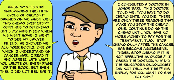
Let me ask you to ponder what Einstein wrote:

1 Insanity-by-Einstein

Do you see any truth is that statement?

Many of us may want to ask: Where is the problem? What causes this problem? What can we do about it?

Jack told us that his oncologist is a very nice man and he was trying his best to cure him. Understandable. We cannot blame the doctor. Legally and medically, apart from chemo or oral drugs, he has nothing else to offer you. He can’t ask you to take supplements or herbs, etc.

Unfortunately, some oncologists will rip apart those who dare to suggest that patients  take herbs, vitamins or control their diet. Nonsense they say — all these non-medical ways are not scientifically proven.

But hang on, is the present day treatment like above  based on “real science”? Granted, these chemo-drugs have undergone clinical trials and have been approved by Government Authorities. But, what does all this mean? The approved drugs can cure your cancer? Far from it! Ask,  why do we see failures after failures being repeated over and over again? It appears that failures  seems to be the norm rather than an exception.

Chemo drugs 3 percetn effective chemo drugs only 25 percent effective

I have one suggestion for those who want to do something!  Think about it seriously.

Granted, chemo-drugs sometimes can make the tumour markers drop to normal level or the tumour shrunk completely after the treatment. But what do you do after this achievement? Send the patient home and ask him to live the same style of life that he/she had before — the earlier life that promoted his/her cancer?

Free fall

  • What if we have a program that teaches patients how to live a healthy life after being effectively treated?
  • What if we teach them to change their life style and diet?
  • What if we ask them to take supplements, vitamins or herbs to make them healthy?
  • What if we ask them to take time to exercise?

These are things that patients can do for themselves when they go home after their “apparently successful” medical treatment.  Above all, these are “harmless” efforts that can result in a better and healthier cancer-free life.

  • In short, why can’t the medical establishment work together with the alternative healers to try and help patients prevent or minimize their cancer recurrence?

At CA Care we teach patients all the above. We take over after patients decide not to go for any more medical treatments. And often for those who are really committed, we succeeded in helping patients to heal themselves.


Surgery, Radiation and Chemo Did Not Cure Malignant Peripheral Nerve Sheath Tumour (Neurofibrosarcoma)

Alex (not real name) is 23 years old. He was born with a certain nerve disorder. His right arm had many benign lumps. When he was 11 years old, his right palm started to swell. He could not write with his right hand anymore.

Composite-1-Hand

Alex underwent a debulking surgery — the lumps were removed. He was told that it was not cancerous. Later in September 2013, there was a swelling in his right arm pit. The mass grew bigger and became painful.

Composite-2-Armpit

A CT scan in January 2014 indicated:

  • A large axillary mass — malignancy needs to be considered. The right subclavian, axillary and brachial arteries pass above and lateral to this mass.
  • No evidence of lung or mediastinal metastasis.

A true-cut biopsy was done and indicated:

  • High grade spindle cell sarcoma favouring malignant peripheral nerve sheath tumour.

Alex underwent an operation to remove this entire right upper limb. Below is the surgeon’s note indicating the extensiveness of the surgery.

Alaister Lim scan

Histopathology confirmed a malignant peripheral nerve sheath tumour (T2bNoMo), Stage 3.

After the surgery Alex received 32 times of radiation treatment. There was no other medication.

Barely six months later, August 2014, the cancer spread to his lungs. Alex underwent 3 cycles of chemotherapy. The treatment was not effective. Alex again had more chemo — 12 cycles in all. The drugs used were Gemzar and Cisplatin.

lung--600

In February 2015, follow up examination showed:

  • Mixed response of the lung nodules. Some nodules are smaller but some previously small ones are larger.
  • A new nodule seen in the left upper lobe adjacent to the left 4th rib.

Chemotherapy did not help control his lung metastasis. Alex decided to give up medical treatment and came to seek our help.

Comment

It is indeed a sad story. As we talked to Alex, we had full admiration for his determined spirit. No, he did not sulk in spite of the fact that he had gone very rough patches since he was born. When he was not able to write with his right hand, he learned to write with his left. He attended university in Australia. When asked if there was anyone taking care of him there, he replied, No, I took care of myself. When medical treatment failed him, he said he wanted to try another way. There was no sign of bitterness in his voice. And he was determined to live.

Internet information about malignant peripheral nerve sheath tumour

Malignant peripheral nerve sheath tumors — MPNSTs (also called neurofibrosarcomas):

  • are a type of cancer that occurs in the protective lining of the nerves that extend from the spinal cord into the body.
  • can occur anywhere in the body, but most often occur in the deep tissue of the arms, legs and trunk.
  • tend to cause pain and weakness in the affected area and may also cause a growing lump or mass.
  • occur more frequently in people with an inherited condition that causes nerve tumors (neurofibromatosis) and in people who have undergone radiation therapy for cancer.

http://www.mayoclinic.org/diseases-conditions/malignant-peripheral-nerve-sheath-tumors/basics/definition/con-20035841

  • are aggressive, locally invasive soft tissue sarcomas, typically presenting as a rapidly growing and painful lump.
  • these tumours account for up to 10% of all soft tissue sarcomas.
  • a common cause of nerve sheath tumors is neurofibromatosis type 1 (NF1).
  • Evans et al. estimate the lifetime risk of developing MPNSTs in the population of patients with NF1 to be as high as 13%.

http://www.hindawi.com/journals/sarcoma/2009/756395/

  • a very rare tumor, with an incidence of 1 per 1,00,000 population.
  • these tumors may arise spontaneously in adult patients, although 5% to 42% of MPNST have an association with multiple neurofibromatosis Type-I

http://www.ncbi.nlm.nih.gov/pmc/articles/PMC1560134/

  • are rare sarcomas with one of the poorest prognoses of all the soft tissue sarcomas.
  • information about adjuvant treatment is scarce.

http://www.ncbi.nlm.nih.gov/pubmed/21303750

  • NF1 is an autosomal dominant condition with a birth incidence of about 1 in 2500 and a prevalence of 1 in 4000.

http://jmg.bmj.com/content/39/5/311.full

  • A peripheral nerve sheath tumor (PNST) is a neoplasm arising from a peripheral nerve.
  • All ages and both sexes may be affected by PNSTs.
  • Sporadic MPNSTs are most common between 40 and 50 years of age, while those occurring in the setting of NF1 are diagnosed some 10 years earlier.

http://atlasgeneticsoncology.org/Tumors/PeriphNervSheatID5094.html

  • Peripheral nerve tumors are uncommon and many neurologists will see only a handful in their careers, unless they have a practice enriched in patients with neurofibromatosis.

http://www.uptodate.com/contents/peripheral-nerve-tumors

  • The role of chemotherapy in advanced malignant peripheral nerve sheath tumor (MPNST) is unclear.

http://annonc.oxfordjournals.org/content/early/2010/07/23/annonc.mdq338.full

  • MPNSTs are highly aggressive in NF1. Conventional chemotherapy does not seem to reduce mortality, and its role must be questioned.

http://www.ojrd.com/content/8/1/127

  • The recommended treatment of this type of sarcoma is the surgical extirpation including wide margins and complementary radiotherapy.However, the prognosis is poor. http://www.sciencedirect.com/science/article/pii/S174194090500004X
  • Malignant peripheral nerve sheath tumors are rare soft tissue sarcomas. They are considered to carry a poor prognosis with current therapeutic approaches.

http://www.casesjournal.com/content/2/1/7612

  • Malignant peripheral nerve sheath tumors (MPNST) are uncommon, biologically aggressive soft tissue sarcomas of neural origin that pose tremendous challenges to effective therapy.
  • Prognosis is generally poor, with high rates of relapse following multimodality therapy in early disease, low response rates to cytotoxic chemotherapy in advanced disease, and propensity for rapid disease progression and high mortality.
  • MPNSTs comprise ∼2% of all sarcomas, a small fraction of a group of cancers that affect 5 people per million per year.
  • Whereas MPNST may arise at any age with no gender predilection, it tends to present earlier in life than most other complex sarcomas.
  • Half of MPNSTs are associated with neurofibromatosis type 1 (NF1), the autosomal dominant condition that, affecting 1 in 3000 live births, represents the most common human cancer genetic predisposition syndrome.
  • The lifetime risk of developing MPNST in a patient with NF1 syndrome is 8%–13%.
  • In general, MPNST is known to have high metastatic potential and poor prognosis.
  • As is the case with all soft tissue sarcomas, complete surgical extirpation with clear margins is the treatment of choice.
  • In the case with most large (>5 cm) high-grade limb sarcomas, adjuvant radiation is advocated to reduce local recurrence. The risk-benefit profile of adjuvant radiation in patients with NF1 must be carefully discussed with all patients in view of the heightened risk of radiation-induced sarcomas.
  • There are no randomized data examining adjuvant chemotherapy specifically in MPNST.

http://theoncologist.alphamedpress.org/content/19/2/193.full


Kidney Cancer Spread to the Lung Six Months After Surgery

SS is a 71-year-old male. Three years ago, in 2012, his urine contained traces of blood. A checkup in a private hospital showed:

  • Normal meatus (urinary meatus is the opening or hole at which urine comes out of the urethra) and urethra (theurethra  is a tube that connects the urinary bladder to the urinary meatus for the flow of fluids from the body. In male  the urethra travels through the penis, and carries semen as well as urine).
  • Mildly enlarged prostate, measures 5.5 x 4.0 x 3.8 cm.
  • Both kidneys are well visualised and shows a normal echo pattern with no focal lesion within it. No renal stones seen and there is no evidence of any hydronephrosis.
  • Creatinine level was elevated, at 156.8 (normal 63.6–110.5).
  • Fatty liver change.

SS was prescribed some medications. The bleeding stopped and he was well for two years.

One day in late 2014,  SS had pains when he turned his body from the right to the left. USG on 9 October 2014 showed:

  • Fatty liver change. No focal liver lesion.
  • A cyst with wall calcifications on the upper pole of the right kidney measuring 8 x 7 mm.
  • There is a lobulated hypoechoic lesion in the upper two thirds of the left kidney measuring 7.0 x 6.8 x 6.2 cm.
  • No hydronephrosis seen.

A CT scan, done on 16 October 2014, showed the following:

  • Small cyst in the upper pole of the right kidney measuring 5 xc 5 mm in size. No mass or focal lesion noted.
  • An irrregularly enhancing mass infiltrating almost the whole of the left kidney sparing the power pole measuring 7.8 x 6.2 x 10.3 cm.
  • Enlarged lymph nodes noted medially measuring 2.5 x 3.0 x 3.0 cm and 1.8 x 1.6 x 1.0 cm.
  • Subcentimeter paracaval and para-aortic lymph nodes.

Comp-Kid-ca


SS underwent surgery to remove his left kidney. The histopathology report dated 21 October 2014 indicated:

  • Grade 3 transitional cell carcinoma of the renal pelvis, T4N2Mx, Stage 4.
  • Two (2/10) hilar lymph nodes contain deposits of metastatic transitional cell carcinoma.
  • Free of metastatic tumour deposits.

The operation was a success (later you would want to ask if this is really true?) and everything was alright. SS was asked to go for chemo, he refused. Routine check up three months later showed everything was perfect. Another routine three-month check up on 22 January 2015 also showed perfect result.

Six months after surgery, 2 June 2015, X-ray showed an oval nodule in the left mid zone of the lung, measuring 13 x 12 mm. This nodule was not seen in the previous chest radiograph on 22 January 2015. An underlying metastatic deposit is considered.

CT scan of the thorax on 4 June 2015 showed:

  • A nodule in the apical segment of the left lower lobe measuring 15 x 12 x 13 mm.
  • Another smaller nodule is noted in the lingular segment of the left upper lobe measuring 5 x 5 x 5 mm.
  • These are suspicious of metastatic deposits.

Composite-Lung-Metastasis

SS was asked to go for chemotherapy. He went to consult an oncologist. Listen to this video.


You want me to do chemo, but I only have one kidney left. How does that affect me?

No problem. We shall use the drugs, carboplatin + Gemzar.

Can you cure me?

No cure. There is no cure for cancer! We shall go for 3 cycles first and see what happen.

Each cycle of chemo is going to cost RM 5,000.

After the chemo (and this is not a cure) how long do I have?

I am not God

I know that you are not God.

Probably you have six months to a year! I can’t say, all depends on yourself!

(Depend on luck? Fong shui? If this being the case, where is the so called “science” in oncology then?)


Lung Cancer and Prostate Problem — Refused chemo & radiation, and still alive?

Aman is a 72-year-old Indonesian male. He and his wife came to seek our help two years ago, April 2013.  He brought along the report of his Whole Body CT scan, dated 21 March 2013, which indicated the following:

  • Spiculated soft tissue mass noted in the right upper lobe of lung, measuring 4.7 x 4.2 cm. The mass extends medially to right hilum and is abutting the adjacent right oblique fissure.
  • No nodule noted in left lung.
  • Enlarged lymph node in precarina region, measuring 1.4 cm.
  • Prostate is enlarged, measuring 5.1 x 6.1 x 5.8 cm with compression effect to urinary bladder.
  • No evidence of distance metastasis in brain and liver.

Composite

Aman was asked to undergo chemotherapy. He refused. Later, he agreed to radiotherapy but could not start the treatment after the machine broke down. Aman then came to seek our help and was started on herbs. We did not get to see Aman again after his first visit.

About two years later, in June 2015, Aman ‘s wife came to our centre and said Aman was doing fine and she wanted some more herbs for her husband.

Here is the gist of our conversation that day (listen to the second half of video below).

Chris:  You (and your husband) first came in 2013 – two years ago. He was scheduled for radiotherapy — did he go for that radiation?

Wife: No, we came here right away — no radiation done. Since then he (husband) never came back to Penang again.

C: In April 2013, he took the herbs. Is he okay now?

W: Yes.

C: Did you ever go back to consult with the doctors again?

W: No.

C: It has been 2 years now and his condition is okay?

W: Yes, okay. Sometime in the morning he has phlegm in the throat. On and off there is a bit of cough.

C: Apart from that, are there any other problem?

W: No problem — every morning he goes for morning walk.

C: Oh, he does not stay home — feeling sick?

W: No, no. He operates a grocery store selling sugar, rice, etc.

C: And he can work?

W: Yes.

C: Amazing indeed. It’s been 2 years . I can’t believe this. (Tell me again) is he okay? No problem, can work?

W: He wakes up early morning and at 6 a.m. and goes for a walk, round and round the neighbourhood. Everyone who sees him says he is healthy!

C: Oh, and he has no other complaints at all?

W: No.

C: His breathing is good?

W: Okay.

C: What is the problem now?

W: No problem.

C: Okay, amazing healing indeed. Continue to do what you are doing!

Comment

This is rather an amazing healing story. How could he survived for that long without chemotherapy or radiotherapy? That is if you believe these treatments are good for cancer! But for us at CA Care, surviving lung cancer for a few years without medical treatments is nothing unusual.

Let’s go back to Aman’s first visit to our centre two years ago and see what transpired during that visit.


Chris: Come, come. You are from Aceh (Indonesia)?

Patient: Yes.

C: You speak Hokkien?

P: Yes.

C: Who is sick?

P: Me, with cancer.

C: What happened? Tell me what happened from the beginning.

Wife:  We went for a checkup in Hospital A. This was in September 2012. They said his lung was “dirty”.

C: He went for a checkup. He had no problem at all? No cough, etc.?

W: Nothing. Actually I went for a checkup. After that he also had a checkup.  The doctor said his lung was “dirty” and his prostate (PSA) was also high, 8.8.

C: Did the doctor give you any medication?

W: Nothing.

C: No medication?

W: No. So we went to Hospital B and did another CT scan. We were told there was a mass. But we went home without doing any treatment.

C: You went to Hospital B. Did another scan.  You knew there was something inside but you did not want any treatment? Now to come and see me — it’s already April 2013 — what did you do from September 2012 till April 2013?

W: We came back to Penang again in January 2013. This time we went to Hospital C. We did another scan.

P: They also took a tissue out — through my back.

C: What did they say?

P: It was cancer.

C: What did you do after that (biopsy)?

P: Nothing.

C: You came to Hsopital A, then Hospital B and then Hospital C. You did one scan after another. Then the biopsy. And you did nothing after all these — what were you trying to do? Okay, in January 2013, after the biopsy you did nothing. Was there any pain? What happened after that.

W: We came back to Penang again in February 2013 and went to a cancer hospital. They gave us an oral medication to take (showing the prescription of Tarceva). But the medicine was not good for him.

C: How long were you on this drug (Tarceva)?

W: After taking 28 tablets (two tablets per day), it was not suitable. We told the doctor about this but he said not to worry and asked us to continue taking the medicine.

C: Why did you say not suitable?

P: Rashes and itchiness (showing hands and face).

C: Whole body?

P: Yes.

C: How much did you pay for this drug?

P: RM 7,500 per month.

C: Before taking this drug (Tarceva) did the doctor not warn you about this side effect?

W: Yes, he did tell us. When we phoned him he told us: Continue. It’s okay. You are reacting to the drug!

C: Did you ask if the drug can cure you?

W: He (husband) did not want chemo injection. So the doctor offered this oral drug instead.

C: After you stopped taking this drug (Tarceva), did you go back to the doctor again?

W: We came back on 21 March 2013 and did a CT scan. They scheduled us for radiotherapy.

C: Did you go for radiotherapy?

W: Not yet. We were supposed to do 30 times. When we turned up for the treatment, we were asked to come back the next day because the machine was not working. Then, the next day, they called to say that the machine was still not working.

C: Have you paid the money for this treatment? How much?

W: Yes, we have already paid the money to the hospital. It was RM 8,000 plus. They told us to come  back again tomorrow to start the treatment.

C: So now, are they going to refund you the  RM 8K that you have you paid?

W: I don’t know.

Advice from CA Care

C: You come here to seek our help. I am going to tell you honestly that I cannot cure your cancer. You must learn how to take care of yourself. Now, you have to stop smoking! Go home and take care of your diet. You cannot eat all those fried foods. No sugar, no diary. No banana for people with lung problems. So you have to take care of your diet. Then take the herbs properly. No cure but these may help you. If you have no problems after this – that’s good enough.

Then don’t be “dumb” anymore — no more going from one hospital to another to take pictures. There are still many more hospitals for you to go to! There is no need to do all these scans. Enough. I can’t cure you but if you are okay — no problem — then don’t ask for more.

For those who want to know more about the “bad and good” stories about lung cancer, read these two comic books.

Lung Cancer: What Now?

Title-Page-Book 1

Lung Cancer Success Stories

Title page Book 2


Breast Cancer: Surgery did not cure. Recurrence and cancer spread extensively to her lungs. But does she want to live?

TTH is a 73-year-old Indonesian lady. In early 2014, she felt a lump in her right breast. If touched she felt pulling pains in her breast. She came to Penang for consultation.

USG on 26 June 2014, indicated a spiculated hypoechoic mass at 11.00 o’clock, 3 cm from nipple. The mass measures 17.7 x 16.3 x 24.0 mm. There are feeder vessels seen supplying this mass lesion. No axillary lymphadenopathy. Impression: Highly suggestive of breast cancer. Category 4.

Breast-USG-S596

A mammogram of both breast also showed similar results.

Chest X-ray showed slight increased reticular markings of both lung parenchyma. No lung nodule seen.

TTH came to seek our advice on 27 June 2014. We suggested that she remove her breast.

Mastectomy

TTH underwent surgery on 7 July 2014.  Histopathology report confirmed:

  • Invasive ductal carcinoma, Stage T1.
  • Resected margins, niple and areola: Free.
  • Right axillary lymph nodes: Sinus histiocytosis (4/4).
  • Tumour negative for estrogen, negative for progesterone and negative for c-erb B2 receptors.

The mastectomy cost RM 6,600. The doctor said no need for chemotherapy or radiotherapy. There was not medication.

TTH came to seek our advise again after the surgery.  We prescribed her some herbs and told her to take care of her diet. We did not get to see THH again after that.

Recurrence and Extensive Lung Metastasis After a Year

A year later, 3 June 2015, TTH and her daughter came to see us again. Why did she come and see us? A routine check up with her doctor on 2 June 2015  indicated extensive lung metastasis. Her CA 15.3 was elevated to 100.2. This time her doctor said she needed chemotherapy and it would be better for her to go home and receive the treatment in Indonesia.

A CT scan done on 2 June 2015 indicated:

  • focal nodule noted in right anterior chest well, on the pectoralis muscle. The nodule measured approximately 8 mm in diameter.
  • There is an adjacent skin nodule approximately 5 mm noted.
  • There are extensive intrapulmonary nodules and masses noted in right and left lungs. They measured approximately 12 to 45 mm n diameter each.
  • Nodular thickening of the interlobular septae noted in both lung bases, consistent with lymphangitis carcinomatosis.

Impresssion:  Local recurrence with extensive lung metastasis and lymphangitis carcinomatosis.

Composite-1

Looking at the above images, we felt real sorry for TTH. How could such extensive metastasis happened within a year? An X-ray done a year ago showed the lung was clear — could this result be wrong?

Chris: You came to see us last year and took some herbs?

Patient:  I took the herbs for a while and stopped. There was nothing wrong with me and I thought I was cured already.

C: Did you take care of your diet?

P: People told me if I don’t eat this and that, I would not have energy – no strength!

C: Your cancer has recurred and it has spread extensively to your lungs. Even if you take the herbs, I am sorry I  cannot cure you.

P: Okay, in that case, there is no need to take the herbs then.

Comment

Last month we posted an article, Breast Cancer: It is all about you — your human nature and attitude

Speaking to TTH, it appeared to us that this patient felt she is already old enough. There is no need to do anything beyond the normal routine that she has been accustomed to. Our experience through the years tells that only 3 out of 10 patients benefited from our herbs. Those who succeeded showed their commitment and wanting to help themselves. Others just came to find an easy way out. Such people are really depressing to work with. It is better that they don’t come and see us at all.

Can the best of medicine in the world cure brain cancer?

This week, there are 3 sad stories about brain cancer.

The Brain Tumor That Killed Beau Biden http://www.thedailybeast.com/articles/2015/05/31/the-brain-tumor-that-killed-beau-biden.html

  • On Saturday, Joseph “Beau” Biden, the son of Vice President Joe Biden and former attorney general of Delaware, died following a long battle against brain cancer.
  • The 46-year-old’s death raises new questions about the disease that killed him—one that’s alarmingly common in adults and chronically misunderstood by the public.
  • This past year there were nearly 70,000 new cases and 14,000 deaths from this disease.
  • There are many different types (of brain cancer) that occur in adults—each with a unique set of features. Each types carries with it a separate prognosis—ranging from a life expectancy of eight months to non-life threatening.
  • Glioblastoma is an aggressive and often fatal tumor, with an estimated 2-year survival rate of around 17 percent for patients between 40 and 65 years old. The underlying cause of GBM is unknown.
  • It’s still unclear which specific type of tumor that Biden suffered from, but whether malignant or benign, it’s clear that the treatment to keep it from coming back ultimately failed.
  • Treatment options typically include a combination of radiation, chemotherapy, and surgery. The goal of surgery is usually to reduce the tumor burden, followed by radiation therapy and certain chemotherapeutics. Chemotherapy may delay the disease course in some cases, but most chemotherapeutic agents for GBM have an extremely low response rate, around 10 percent. The prognosis is not particularly good for any age group.

Brain cancers like Beau Biden’s kill about 15,000 adults each year http://www.washingtonpost.com/news/to-your-health/wp/2015/05/31/brain-cancers-like-beau-bidens-kill-about-15000-adults-each-year/

  • A variety of malignant brain and central nervous system tumors kill about 15,000 people in the United States each year, often after difficult courses of surgery, radiation and chemotherapy.
  • Beau Biden was first diagnosed in August 2013 and after surgery, radiation and chemotherapy returned to normal duties in November that year. But his cancer recurred this spring, and he began treatment again at Walter Reed National Military Medical Center.
  • Former Massachusetts senator Edward M. Kennedy died in August 2009, 15 months after he suffered a seizure and was diagnosed with a malignant glioma, the most common form of brain cancer.
  • For most incurable brain tumors, the primary challenge is managing the portions of the tumor that are undetectable by diagnostic imaging, These tumor cells are often the source of recurrence, traveling undetected to other brain regions of the patient and then driving the formation of new areas of growth with little warning. Most malignant brain tumors eventually recur.

Beau Biden fought bravely before losing battle with brain cancer; here are the lessons you should learn  http://www.nydailynews.com/life-style/health/doctor-lessons-beau-biden-death-article-1.2242753

  • Beau Biden had been dealing with health problems since 2010, when he suffered from a stroke.
  • Three years later … after he suddenly felt disorientated and weak during a vacation, he was admitted to M.D. Anderson Cancer Center in Houston. Not long after that, he was diagnosed with brain cancer. He underwent surgery and was free of the cancer; however, it returned this spring.
  • Biden underwent aggressive treatmentat Walter Reed National Military Medical Center in Bethesda, Md.
  • We do not know what causes brain cancer. Most brain tumors are not linked to any known risk factors. While there is no proof that radiation causes cancer, we do know that radiation is not good for you. The use of cell phones has long been led to debate about whether they may increase a person’s risk for brain cancer. Current research shows conflicting results. Regardless, it is probably a good idea to limit cell phone use as much as possible or at least use a hands-free headset to reduce your exposure to radiation.

Beau Biden Dies at 46 From Brain Cancer: Why the Disease Is So Hard to Beat https://www.yahoo.com/health/beau-biden-dies-at-46-from-brain-cancer-why-the-120362278807.html 

  • Details about Biden’s healthprior to his death are scarce. What is known is that he was hospitalized the week up until his death at Walter Reed National Military Medical Center, and that he did have a small lesion removed from his brain in 2013 at University of Texas M.D. Anderson Cancer Center, but was given a clean bill of health after the procedure.
  • …. brain tumor is known to have a high propensity for returning, even after being successfully removed
  • No matter what, almost all the time you’re talking about a kind of tumor that is going to come back and become more resistant to therapies,” Weiss, who specializes in treating adult malignant and benign brain tumors, tells Yahoo Health. “That’s why you hear these patients have a brain tumor, a primary brain tumor, and they’ve been given a clean bill of health. But they’re never really in a position” to have a clean bill of health for the rest of their lives, she says.

Case from Singapore

Someone from Singapore sent me this message:

Dear Doctor Chris,

Thank you for reading this message! I know it is hard for u to make any diagnosis without seeing the patient – but can I just ask before I bring my 32 year old wife over from Singapore if you are able to help her treat Glioblatoma Multiforme Grade 4 brain cancer. She has done surgery to remove 70% of tumor on 28 April and has not started on Chemo/ Radiation. We hope to let her do Immmunotherapy (T cell) before bringing her to u for treatment – do u advise that and know of a reliable hospital/ clinic in Malaysia for recommendation? Please for your kind advice!

Reply: I have bad impression of IT!

Can we seek your treatment for glioblastoma? … can we please seek your help for her glioblastoma?

Reply: Unfortunately … no cure from that ca

Can extend? For how long?

 Reply: Can’t say or predict. 

Case from Penang

May 30, 2015 at 4:20 PM,

Dear Dr Chris Teo,

My sister is a cancer patient with brain tumors, grade 4, high grade glioblastoma.

Sep 2013- She fainted and was sent to hospital, scan showed she had some bleeding in her brain.

Oct 2013- another scan showed that she had a 3cm big tumor.

She had her tumors removed in Oct 2013 by X of General Hospital Penang due to emergency.

Oct 2014-her tumors grew back and scan report found that there’re 5 tumors.

She had undergone 2 sessions of radiotherapy and 3 sessions of chemotherapy since then.

There is no more chemotherapy for her. Her brain swell every week and had to go for dripping and treat with mannitol.

She has strong will to survive as she has a 4 year old daughter. Please save her. We would like to make an appointment with you to see if there’s any advice from you.

I look forward to your reply. Warm regards.

Reply: I am sorry I cannot save her. Her cancer is very, very difficult.

Dear Dr Chris Teo,

Thank you for your reply.

Although it looks like her condition is very critical but she has very good blood test report, all her other organs are functioning very well.

Other than her speech problem and body coordination due to her brain’s tumors, she has clear mind, strong surviving will, she eats very well and pass motions and urine as normal person.

I just feel that if you’ve any herbs that can help to alleviate the swell and cancer cells in her brain, that would be very helpful.

My mother and her husband are there at your clinic now to seek your opinion. Please at least give them some advice.  My mother has been very careful in her diet … It was very surprise to see a glioblastoma cancer patient to survive more than 5 months. My sister has already passed her 1.5 years after diagnosed.

This 29-year-old patient (on wheel chair), her husband and mother came to see us. The following is a letter from her oncologist:

Mdm H is my patient since 2014 with brain cancer, now recurrence with multiple nodules in the brain. This has left her with weakness of her limbs, unsteady gait and double vision. She is therefore dependent and reliant on her carers for mobilising and self-care at home. Her disability is deemed permanent in view of her recurrence of cancer in the brain. She gets agitated and irritated due to her tumour in the brain. Hope you will understand the emotional changes that she is going through due to her illness.

Composite-1

Mdm H underwent surgery on 18 October 2013. This was followed by 30 sessions of radiation and 2 cycles of chemotherapy given as 12 injections. The drug used was Avastin.

By 2 September 2014, H was told that there was NO MORE cancer! Her mother said H was cured!

Unfortunately, about 2 months after this “great news” the cancer came back again. H again underwent chemotherapy. After the 11th injection, the oncologist in a private hospital told her to go and seek further treatment in a government hospital.  As far as he is concerned he could not help H anymore.

At this point, the family decided to come to us for help. I told the family. I cannot cure your cancer. 6 Surgery Tobias Brain ca

It is indeed sad that patients and their families don’t seem to understand that surgery does not cure any cancer (unless it is of a very early stage).  Listen to this: 5 Ssurgery come back again

Over the years, I have come across many cases of brain cancer. Here are some of their stories:

Brain Tumour: Operation, you have an 80 percent chance of cure! Do you believe that?

Brain Cancer: Surgery and Radiotherapy. Died

Brain Cancer: Radiotherapy – Recurrence; Chemo – Recurrence; and Avastin – Dead

Helping a Six-Year-Old with Cancer of the Brain Stem

Brain Tumour: Herbs and e-Therapy Helped Him

Brain Cancer – Marvelous Technology, Dismal Outcome

Brain Cancer – Yet She Lives !

Brain Cancer: She Was in a Coma

Declined Surgery Only On Herbs

Brain Tumour Shrunk With Herbs


Look at the Bigger Picture Part 2. You don’t have to “swallow” everything that they offer you!

Look at the Bigger Picture Part 1. Misguided advice about your diet!

Ina called to ask if she could come to our house. She was so excited and wanted to share with us the “good” news after meeting her doctor.

Who is Ina?  She is our patient and also a friend. She was diagnosed with breast cancer in August 2012,  almost 3 years ago. She had a mastectomy but declined chemotherapy, radiotherapy and taking of Tamoxifen. Indeed a daring lady!

Ever since after her surgery to this day, Ina had been under pressure from her doctor urging her to go for chemo and radiation. Ina flatly refused! Then the doctor insisted that at least she should take Tamoxifen!

Why was Ina so excited and wanting to see us?  Two reasons.

Reason number two.  Her liver was just doing fine! Now, her doctor did not bring up the subject of her liver metastasis and this time did not “pushed” her to go for chemotherapy!

A misdiagnosed liver metastasis?

For the past two years Ina had been living under stress. After being diagnosed with breast cancer in August 2012 and had a mastectomy, her doctor insisted that she undergo chemotherapy.  Her doctor was unhappy that Ina was adamant not to follow his advice! In fact, the doctor was also not happy with a Chris Teo for not pushing Ina to go for chemo!

Ina’s doctor became more unhappy after the result of an USG on 20 January 2013. The doctor told Ina that her cancer had spread to her liver based on the result below:

A few hypoechoic cysts measuring 9.6 to 18.4 mm are seen in the right and left lobe of liver. No solid component is seen within these cysts. No other focal liver lesion is seen.

Ina was asked to go for a PET scan, and an X-ray. Ina refused these procedures.

Then, about a year later, a follow up USG showed the following:

  • The right lobe of liver shows mildly hyperechoic module close to the IVC and the medial part of the right hemidiaphragm. This measures about 38 x 27 x 36 mm. Apart from this, there is at least another mildly hyperechoic solid nodule in the right lobe in segment 8, measuring about 12 mm.
  • Smaller nodules cannot be excluded in the right lobes of the liver.
  • There are two simple cysts in the right and left lobes of the liver, measuring about 25 and 12 mm.

Impression: In view of past history, I would suggest further imaging to exclude metastatic disease. Haemangionmas may also give a similar appearance.

The doctor insisted that Ina go for a PET scan! Ina again refused. The doctor asked Ina, Don’t you tell Chris Teo that the cancer has already gone to the liver?  The doctor wrote Chris Teo a letter below!

Select-Ed-600

It is nice of her doctor to caution me with this note. But yes, I was very much aware that breast cancer can spread to the liver, bone, lung or the brain. I have also seen cases after cases of metastatic cancer before (agreed, not as many as the doctors!). Let me remind you what I wrote in my book, Your Breast (pg.120, 135 and 139).

Pg 120 Pg 135 Pg 139

The questions we posed to Ina were basically this:

  1. Okay, you were asked to do the PET scan – to know if the cancer had spread to any other parts of the body. But do you want to follow up with medical treatments. Go for chemo or radiation or even surgery again if there was a spread to somewhere? Ina said, NO – I would not want any more medical treatments. That being the case, why go for a PET scan then? What benefit do you get from just wanting to know?
  2. Now, the doctor implied said that there were secondaries in her liver. Ina needed chemotherapy right away. Ask the doctor first, Can chemotherapy cure her metastatic liver cancer?

Before her death, Amy Cohen Soscia – a breast cancer patient in the US, said: There is no cure for metastatic breast cancer. It never goes away. You just move from treatment to treatment.

Was Ina’s liver metastasis a misdiagnosis?

In May 2015, we requested Ina to go for a checkup since at that time she was concerned about a lump in her right breast (Part 1 of this story).  USG of the abdomen was done and the following were the results.

  • The right lobe of the liver shows a simple cyst measuring about 28 mm. There are two mildly hyperechoic solid right lobe hepatic nodules which are probably haemangiomas. They have not changed significantly in sizes since the previous examination.
  • The larger lesion seen near the IVC measures about 37 x 28 x 37 mm. The smaller lesion seen more peripherally measures about 9 mm in the right lobe of the liver.
  • The left lobe of the liver also shows a small cyst measuring about 9.8 mm in size.

The results of Ina’s liver function test were as follows:

Liver function
ALT 15
AST 23
GGT 19
Alkaline phosphatase 49


Based on the above results, the doctor “stopped” talking about Ina’s liver problem. The question of her undergoing chemotherapy for her liver metastasis just vanished! Ina was very much at ease. So happy — NO more pressure from her doctor!

After about 3 years later, someone has decided that the “perceived” liver metastasis is probably not a cancer after all. The question which I would like ask is this, What could have happened to Ina if she were to follow her doctor’s advice and went for chemotherapy for the “perceived” liver metastasis? Would she be as healthy as she is today or dead? Your guess would be as good as ours!

Bravo, after taking the liver herbs the results were clear. The liver nodules have not changed significantly in sizes since the previous examination. If the herbs were not helpful, the nodules would have grown bigger. Right? No? And more important, if the herbs were useless, Ina would have been very sick by now! No?

We always tell patients to learn to live with their cancer! We praise God for this blessing.

Ina’s liver nodules are not giving her any problem – and she is well, healthy with no pain; can eat, can sleep and can go about doing her normal daily routine – what else does she want? That is looking at the bigger picture! She need NOT do things to please her doctor!

As a gentle reminder, can you learn something from these two sad stories? Or do we have to write more of such stories to make you understand?

2.3 cm Malignant Breast Lump: Surgery, Chemo and Radiation — Disaster

https://cancercaremalaysia.com/2015/04/25/2-3-cm-malignant-breast-lump-surgery-chemo-and-radiation-disaster/

Selling Empty Promise ?: https://cancercaremalaysia.com/2014/05/21/breast-cancer-part-23-does-chemotherapy-make-sense/

empty-1empty2empty3empty4empty5empty6

 (The above failed treatment in Singapore cost about RM 500,000)

Look at the Bigger Picture Part 1. Misguided advice about your diet!

Ina called to ask if she could come to our house. She was so excited and wanted to share with us the “good” news after meeting her doctor.

Who is Ina?

She is our patient and also a friend. She was diagnosed with breast cancer in August 2012, almost 3 years ago. She had a mastectomy but declined chemotherapy, radiotherapy and taking of Tamoxifen. Indeed a daring lady!

Ever since after her breast surgery to this day Ina had been under pressure from her doctor urging her to   go for follow up chemo and radiation. Ina flatly refused! Then the doctor insisted that at least she should take Tamoxifen!

Why was Ina so adamant in not wanting to follow her doctor’s advice?  

Before her breast cancer diagnosis, her mother had lung cancer.  She brought her mom to see us and was on the herbs. So Ina knew what the herbs could do! Compare that to her father who had suffered and died of cancer, at least mom was much better off with the herbs. Also, Ina (being a business woman) knows of many people who have cancer and how they suffered while undergoing chemotherapy! Some died.

Ina’s sister had lumps in her breast and had to undergo surgery a few times to remove these lumps. But after taking our herbs, she said “good bye” to her routine breast lump surgery!

Probably all these made Ina realised that there is “another way” for her.  She decided to “stick” with us.

Why was Ina so excited and wanting to see us?  

Two reasons. Reason number one — No sign of recurrence and right breast lumps were benign.

A few days before seeing her doctor, Ina came to see us. She felt a lump in her right breast. And this bothered her very much. It hurts when she lifted her right arm. As usual, our advice was, Don’t panic!  Go and see your doctor and do an USG. Find out what exactly it is. And then come back and we discuss what to do after that.  We cautioned her:  Of course, if it bothers you then have the lump removed. At the same time, do a full blood test.

Results of the blood test: 14 May 2015

Total WBC 4.2
Total RBC 4.0
Haemoglobin 12.1
Platelet count 192
ESR 7
Creatinine 66
Tumour markers
CA 125 9
CA 15.3 17.10
CA 19.9 16.66

Composite-1

USG of right breast: 14 May 2015

  • Multiple right breast simple cysts, some of them containing inspissated fluid. The largest is a palpable lesion close to the areolar margin and at around 8 o’clock position measuring 31 mm.
  • The second measures about 7.5 mm at 3 o’clock position.
  • The others measure about 3 to 7 mm in sizes.
  • No solid right breast mass seen.
  • A benign right axillary lymph node seen measuring about 15 x 6 mm.

Based on the above report, Ina’s doctor said she is healthy and there is no cancer. Of course, like all patients, Ina was happy to hear such a positive comment. BUT, this good word was followed by a depressing statement. The doctor said, But you look thin. You must been following a special diet. No, go on and eat anything you like. Are you still on Chris Teo’s herbs?  Ina answered, YES, absolutely.

The doctor wanted to remove the big lump in her right breast. Right away, he scheduled a surgery for Wednesday — a few days away. Ina said she wanted to go home and think about it first.

When Ina told us about this, we reminded her that even before her cancer diagnosis, she already had 6 lumps in her right breast (only the lump in her left breast was malignant). It hurts when she lifted her right arm and the pain was along her bra line.

It hurts when you wear your bra? The answer is Yes. What if you don’t wear bra? Does it hurt? No! We suggest that Ina stop wearing her bra!

Another question we posed was, The doctor wanted to remove only one lump in the breast. What about the rest (5 others)? Wait for them to grow bigger and then do surgery again and again like her sister?

What if Ina were to take the herbs for breast lump for about 3 months and see what happens?

Ultrasound 2012

Indeed if we compare the two reports, we know that 3 years ago the largest cyst was already 2.9 cm. Perhaps this one has gown bigger. After our discussion Ina decided to defer her surgery!

Eat anything you like and grow fat!

What is most upsetting to Ina is her doctor’s insistence that she go ahead and eat anything she likes. He has been harping on this issue every time Ina went for her routine checkup (another more annoying and dangerous insistence is the need to undergo chemotherapy or taking Tamoxifen — another story to follow).

This is our message to all cancer patients

Diet plays a significant role in your healing of cancer. It plays a similar role in the recurrence of your cancer. Take care of your diet.

Is our advice on diet based on scientific evidence? Of course, there are thousands of papers in medical journals about this “sore and sick” topic on diet and cancer. What you need to do is just take time to read them. Get yourself educated. If you don’t have the time to read, at least take a few minutes to “listen” to what doctors themselves have got to say.

2-Medical-profession-which-

3 Oncologist-dont-know-nutrit

1 deVita

10  Nutriton-stop-growth-spread

8-Diet-must-be-integral-par

6 Pig-knows-better-nutrtion

Capturefood 2

Capturefood

In this article, we asked you to look at the bigger picture. Is healing of cancer only about surgery, chemo, radiation or taking of Tamoxifen?

There is another point – make a google search using this phrase, wearing of tight bra and incidence of breast cancer. You will be surprised to find articles about this subject which your doctors may think it is just another “crazy” idea.

This is what I got after my google search.

Bra-and-cancer

In the “Bra and Breast Cancer Study” in the United States, it was discovered that women with breast cancer had a history of sporting tighter and longer bra-wearing than did the women who had not (yet) developed the disease. In fact, virtually all of the cancer group wore bras over 12 hours daily with 18% even sleeping with their bras which means they wore bras 24 hours daily. In contrast, the comparison groups had 25% of the women either bra-free or wearing bras less than 12 hours daily.  Only 3% of this group slept with their bras on. This study revealed a significant link between bras and breast cancer that is three times greater than the link between cigarette smoking and lung cancer! http://www.health2us.com/bra.htm

Women everywhere are discovering that wearing bras can make their breasts droopy and stretched out, and also cause cysts, pain, and cancer. For some women, enough said. The bra goes. It was the first thing they took off after work, anyway. It was always so uncomfortable. And more women are becoming bra-free in the name of comfort and health. For other women, no way! The bra stays no matter what. The cancer detection and treatment industry loves these women. They want women to wear bras. With one million bras sold EACH DAY in the US alone, that’s a lot of women binding and constricting the health out of their breasts in the name of fashion. http://www.killerculture.com/breast-cancer-is-preventable/

As a breast cancer surgeon, I not infrequently have to deal with many of the common myths that have sprung up around breast cancer. Some are promoted by quacks; others are just myths that sound plausible but aren’t true … One such myth has been …. that wearing bras increases the risk of breast cancer is one of those unsinkable rubber ducks (as James Randi would put it) of a myth that just won’t die … It’s unclear where and how long ago this myth first appeared, but there’s little doubt on when it was first popularized: 1995. That was the year that a book by Sydney Ross Singer and Soma Grismaijer entitled Dressed to Kill: The Link Between Breast Cancer and Bras was published. The central thesis of the book was that bra-wearing is a major cause of breast cancer because of its claimed effect on lymphatic circulation. Basically, the idea was (and still is) that bras interfered with lymphatic drainage and thereby, though unclear mechanisms, caused cancer. The claim was that there are all sorts of “toxins” (of course) that cause cancer and that the lymph vessels drain those “toxins” away from the breast. Thus, if you believe Singer and Grismaijer, these “toxins” are concentrated in the breast by the constriction that bras produce and result in breast cancer. https://www.sciencebasedmedicine.org/one-more-time-no-wearing-a-bra-does-not-cause-breast-cancer/

Okay, here we are again. Another great debate like diet and cancer! We leave it up to you to read more and find your own “truth”.

Ina said she had pains when lifting her arms because of her tight bra. She did not have this problem if she did not wear her bra. Our simple suggestion is, either wear a loose bra or don’t wear it at all! For the business minded, why not design a breast-cancer-friendly bra. That may probably cool down the debate a bit and at the same time may enrich your bank account!